Bluetongue virus capsid protein VP5 perforates membranes at low endosomal pH during viral entry.
Xia, X., Wu, W., Cui, Y., Roy, P., Zhou, Z.H.(2021) Nat Microbiol 6: 1424-1432
- PubMed: 34702979
- DOI: https://doi.org/10.1038/s41564-021-00988-8
- Primary Citation of Related Structures:
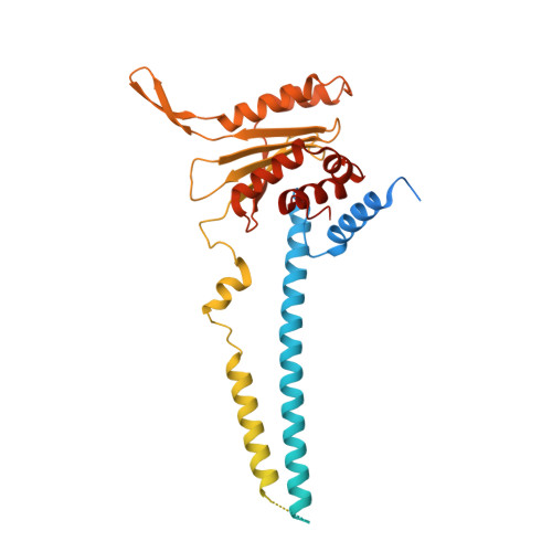
7RTN, 7RTO - PubMed Abstract:
Bluetongue virus (BTV) is a non-enveloped virus and causes substantial morbidity and mortality in ruminants such as sheep. Fashioning a receptor-binding protein (VP2) and a membrane penetration protein (VP5) on the surface, BTV releases its genome-containing core (VP3 and VP7) into the host cell cytosol after perforation of the endosomal membrane. Unlike enveloped ones, the entry mechanisms of non-enveloped viruses into host cells remain poorly understood. Here we applied single-particle cryo-electron microscopy, cryo-electron tomography and structure-guided functional assays to characterize intermediate states of BTV cell entry in endosomes. Four structures of BTV at the resolution range of 3.4-3.9 Å show the different stages of structural rearrangement of capsid proteins on exposure to low pH, including conformational changes of VP5, stepwise detachment of VP2 and a small shift of VP7. In detail, sensing of the low-pH condition by the VP5 anchor domain triggers three major VP5 actions: projecting the hidden dagger domain, converting a surface loop to a protonated β-hairpin that anchors VP5 to the core and stepwise refolding of the unfurling domains into a six-helix stalk. Cryo-electron tomography structures of BTV interacting with liposomes show a length decrease of the VP5 stalk from 19.5 to 15.5 nm after its insertion into the membrane. Our structures, functional assays and structure-guided mutagenesis experiments combined indicate that this stalk, along with dagger domain and the WHXL motif, creates a single pore through the endosomal membrane that enables the viral core to enter the cytosol. Our study unveils the detailed mechanisms of BTV membrane penetration and showcases general methods to study cell entry of other non-enveloped viruses.
Organizational Affiliation:
Department of Microbiology, Immunology & Molecular Genetics, University of California, Los Angeles, Los Angeles, CA, USA.