7RMS

Crystal structure of [I11G]cycloviolacin O2


Experimental Data Snapshot

  • Method: X-RAY DIFFRACTION
  • Resolution: 1.10 Å
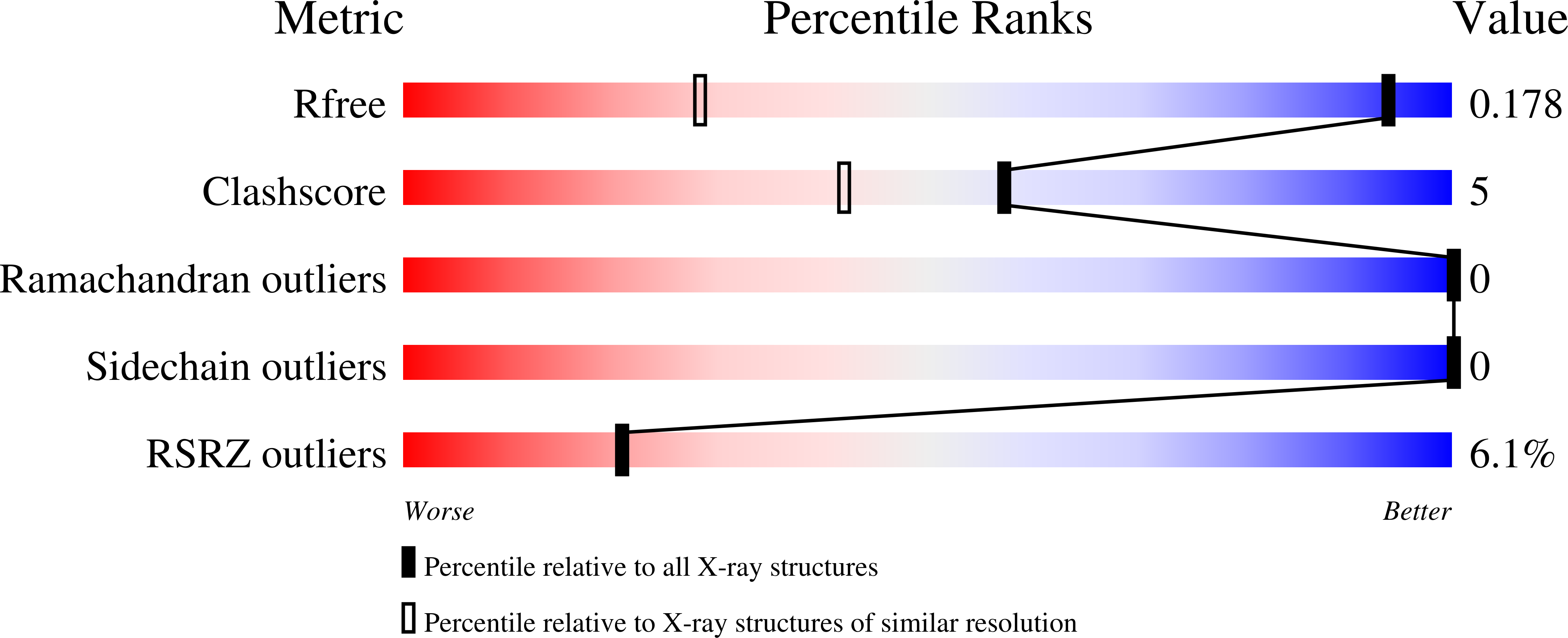
  • R-Value Free: 0.177 
  • R-Value Work: 0.153 
  • R-Value Observed: 0.156 

wwPDB Validation   3D Report Full Report


This is version 1.4 of the entry. See complete history


Literature

Enabling Efficient Folding and High-Resolution Crystallographic Analysis of Bracelet Cyclotides.

Huang, Y.H.Du, Q.Jiang, Z.King, G.J.Collins, B.M.Wang, C.K.Craik, D.J.

(2021) Molecules 26

  • DOI: https://doi.org/10.3390/molecules26185554
  • Primary Citation of Related Structures:  
    7RMQ, 7RMR, 7RMS

  • PubMed Abstract: 

    Cyclotides have attracted great interest as drug design scaffolds because of their unique cyclic cystine knotted topology. They are classified into three subfamilies, among which the bracelet subfamily represents the majority and comprises the most bioactive cyclotides, but are the most poorly utilized in drug design applications. A long-standing challenge has been the very low in vitro folding yields of bracelets, hampering efforts to characterize their structures and activities. Herein, we report substantial increases in bracelet folding yields enabled by a single point mutation of residue Ile-11 to Leu or Gly. We applied this discovery to synthesize mirror image enantiomers and used quasi-racemic crystallography to elucidate the first crystal structures of bracelet cyclotides. This study provides a facile strategy to produce bracelet cyclotides, leading to a general method to easily access their atomic resolution structures and providing a basis for development of biotechnological applications.


  • Organizational Affiliation

    Institute for Molecular Bioscience, The University of Queensland, Brisbane, QLD 4072, Australia.


Macromolecules
Find similar proteins by:  (by identity cutoff)  |  3D Structure
Entity ID: 1
MoleculeChains Sequence LengthOrganismDetailsImage
[I11L]cycloviolacin O230Viola odorataMutation(s): 1 
UniProt
Find proteins for P58434 (Viola odorata)
Explore P58434 
Go to UniProtKB:  P58434
Entity Groups  
Sequence Clusters30% Identity50% Identity70% Identity90% Identity95% Identity100% Identity
UniProt GroupP58434
Sequence Annotations
Expand
  • Reference Sequence
Find similar proteins by:  (by identity cutoff)  |  3D Structure
Entity ID: 2
MoleculeChains Sequence LengthOrganismDetailsImage
D-[I11L]cycloviolacin O230Viola odorataMutation(s): 0 
UniProt
Find proteins for P58434 (Viola odorata)
Explore P58434 
Go to UniProtKB:  P58434
Entity Groups  
UniProt GroupP58434
Sequence Annotations
Expand
  • Reference Sequence
Experimental Data & Validation

Experimental Data

  • Method: X-RAY DIFFRACTION
  • Resolution: 1.10 Å
  • R-Value Free: 0.177 
  • R-Value Work: 0.153 
  • R-Value Observed: 0.156 
  • Space Group: P 1 21 1
Unit Cell:
Length ( Å )Angle ( ˚ )
a = 27.291α = 90
b = 25.108β = 115.72
c = 29.927γ = 90
Software Package:
Software NamePurpose
PHENIXrefinement
PDB_EXTRACTdata extraction
XDSdata reduction
Aimlessdata scaling
PHASERphasing

Structure Validation

View Full Validation Report


Entry History & Funding Information

Deposition Data


Funding OrganizationLocationGrant Number
Australian Science and Industry Endowment Fund (SIEF)AustraliaCE200100012

Revision History  (Full details and data files)

  • Version 1.0: 2021-09-22
    Type: Initial release
  • Version 1.1: 2021-09-29
    Changes: Database references
  • Version 1.2: 2021-10-13
    Changes: Database references
  • Version 1.3: 2023-10-18
    Changes: Data collection, Refinement description
  • Version 1.4: 2023-11-15
    Changes: Data collection