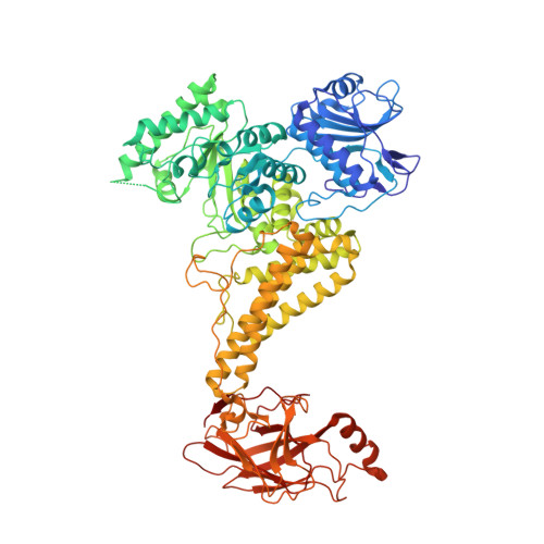
A GH115 alpha-glucuronidase structure reveals dimerization-mediated substrate binding and a proton wire potentially important for catalysis.
Wilkens, C., Vuillemin, M., Pilgaard, B., Polikarpov, I., Morth, J.P.(2022) Acta Crystallogr D Struct Biol 78: 658-668
- PubMed: 35503213
- DOI: https://doi.org/10.1107/S2059798322003527
- Primary Citation of Related Structures:
7PUG, 7PXQ - PubMed Abstract:
Xylan is a major constituent of plant cell walls and is a potential source of biomaterials, and the derived oligosaccharides have been shown to have prebiotic effects. Xylans can be highly substituted with different sugar moieties, which pose steric hindrance to the xylanases that catalyse the hydrolysis of the xylan backbone. One such substituent is α-D-glucuronic acid, which is linked to the O2' position of the β-1,4 D-xylopyranoses composing the main chain of xylans. The xylan-specific α-glucuronidases from glycoside hydrolase family 115 (GH115) specifically catalyse the removal of α-D-glucuronic acid (GlcA) or methylated GlcA (MeGlcA). Here, the molecular basis by which the bacterial GH115 member wtsAgu115A interacts with the main chain of xylan and the indirect involvement of divalent ions in the formation of the Michaelis-Menten complex are described. A crystal structure at 2.65 Å resolution of wtsAgu115A originating from a metagenome from an anaerobic digester fed with wastewater treatment sludge was determined in complex with xylohexaose, and Asp303 was identified as the likely general acid. The residue acting as the general base could not be identified. However, a proton wire connecting the active site to the metal site was observed and hence a previous hypothesis suggesting a Grotthuss-like mechanism cannot be rejected. Only a single molecule was found in the asymmetric unit. However, wtsAgu115A forms a dimer with a symmetry-related molecule in the crystal lattice. The xylohexaose moieties of the xylohexaose are recognized by residues from both protomers, thus creating a xylohexaose recognition site at the dimer interface. The dimer was confirmed by analytical size-exclusion chromatography in solution. Kinetic analysis with aldouronic acids resulted in a Hill coefficient of greater than 2, suggesting cooperativity between the two binding sites. Three Ca 2+ ions were identified in the wtsAgu115A structures. One Ca 2+ ion is of particular interest as it is coordinated by the residues of the loops that also interact with the substrate. Activity studies showed that the presence of Mg 2+ or Mn 2+ resulted in a higher activity towards aldouronic acids, while the less restrictive coordination geometry of Ca 2+ resulted in a decrease in activity.
Organizational Affiliation:
Department of Biotechnology and Biomedicine, Technical University of Denmark, Søltofts Plads 224, 2800 Kongens Lyngby, Denmark.