Reactions with Proteins of Three Novel Anticancer Platinum(II) Complexes Bearing N-Heterocyclic Ligands.
Sacco, F., Tarchi, M., Ferraro, G., Merlino, A., Facchetti, G., Rimoldi, I., Messori, L., Massai, L.(2021) Int J Mol Sci 22
- PubMed: 34638887
- DOI: https://doi.org/10.3390/ijms221910551
- Primary Citation of Related Structures:
7PNH, 7PNI - PubMed Abstract:
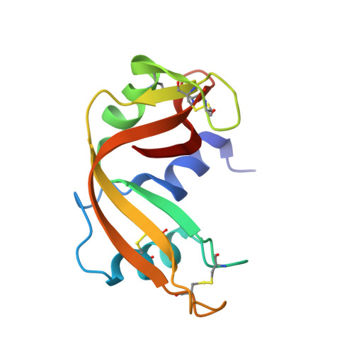
Three novel platinum(II) complexes bearing N-heterocyclic ligands, i.e., Pt2c, Pt-IV and Pt-VIII, were previously prepared and characterized. They manifested promising in vitro anticancer properties associated with non-conventional modes of action. To gain further mechanistic insight, we have explored here the reactions of these Pt compounds with a few model proteins, i.e., hen egg white lysozyme (HEWL), bovine pancreatic ribonuclease (RNase A), horse heart cytochrome c (Cyt-c) and human serum albumin (HSA), primarily through ESI MS analysis. Characteristic and variegate patterns of reactivity were highlighted in the various cases that appear to depend both on the nature of the Pt complex and of the interacting protein. The protein-bound Pt fragments were identified. In the case of the complex Pt2c, the adducts formed upon reaction with HEWL and RNase A were further characterized by solving the respective crystal structures: this allowed us to determine the exact location of the various Pt binding sites. The implications of the obtained results are discussed in relation to the possible mechanisms of action of these innovative anticancer Pt complexes.
Organizational Affiliation:
Department of Chemistry, University of Florence, Via della Lastruccia 3-13, 50019 Sesto Fiorentino, Italy.