7PIV

Active Melanocortin-4 receptor (MC4R)- Gs protein complex bound to agonist NDP-alpha-MSH at 2.86 A resolution.


Experimental Data Snapshot

  • Method: ELECTRON MICROSCOPY
  • Resolution: 2.86 Å
  • Aggregation State: PARTICLE 
  • Reconstruction Method: SINGLE PARTICLE 

wwPDB Validation   3D Report Full Report


This is version 1.0 of the entry. See complete history


Literature

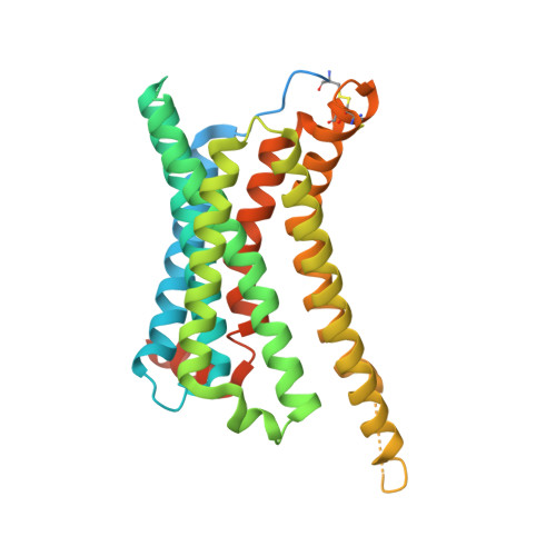
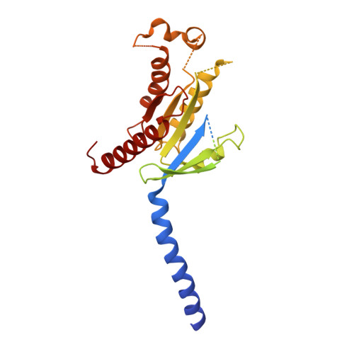
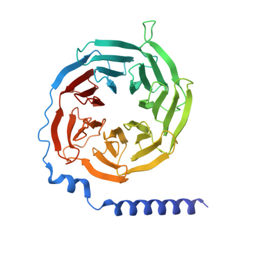
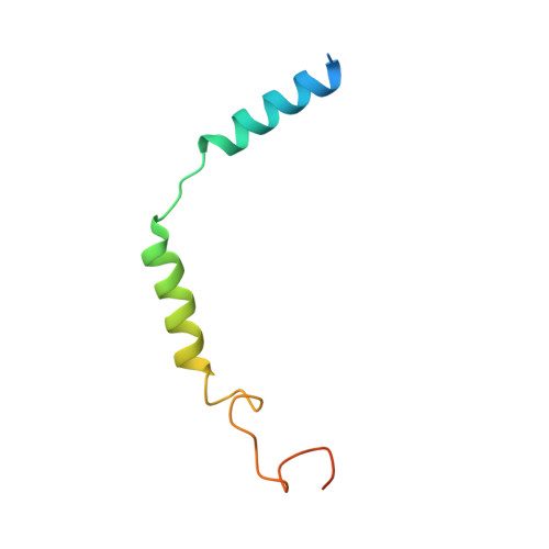
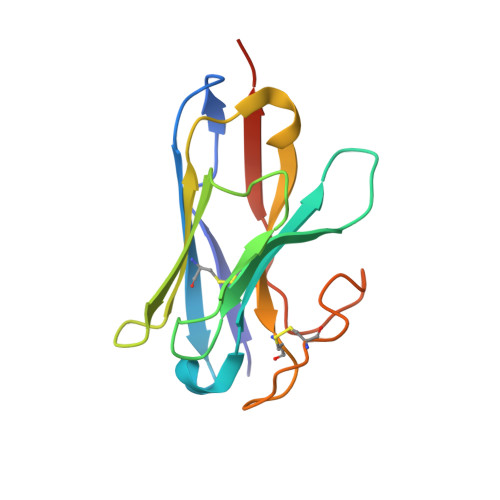
Structures of active melanocortin-4 receptor-Gs-protein complexes with NDP-alpha-MSH and setmelanotide.

Heyder, N.A.Kleinau, G.Speck, D.Schmidt, A.Paisdzior, S.Szczepek, M.Bauer, B.Koch, A.Gallandi, M.Kwiatkowski, D.Burger, J.Mielke, T.Beck-Sickinger, A.G.Hildebrand, P.W.Spahn, C.M.T.Hilger, D.Schacherl, M.Biebermann, H.Hilal, T.Kuhnen, P.Kobilka, B.K.Scheerer, P.

(2021) Cell Res 31: 1176-1189

  • DOI: https://doi.org/10.1038/s41422-021-00569-8
  • Primary Citation of Related Structures:  
    7PIU, 7PIV

  • PubMed Abstract: 

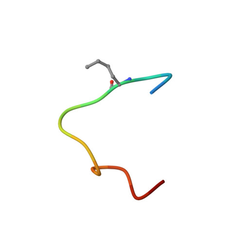
    The melanocortin-4 receptor (MC4R), a hypothalamic master regulator of energy homeostasis and appetite, is a class A G-protein-coupled receptor and a prime target for the pharmacological treatment of obesity. Here, we present cryo-electron microscopy structures of MC4R-Gs-protein complexes with two drugs recently approved by the FDA, the peptide agonists NDP-α-MSH and setmelanotide, with 2.9 Å and 2.6 Å resolution. Together with signaling data from structure-derived MC4R mutants, the complex structures reveal the agonist-induced origin of transmembrane helix (TM) 6-regulated receptor activation. The ligand-binding modes of NDP-α-MSH, a high-affinity linear variant of the endogenous agonist α-MSH, and setmelanotide, a cyclic anti-obesity drug with biased signaling toward Gq/11, underline the key role of TM3 in ligand-specific interactions and of calcium ion as a ligand-adaptable cofactor. The agonist-specific TM3 interplay subsequently impacts receptor-Gs-protein interfaces at intracellular loop 2, which also regulates the G-protein coupling profile of this promiscuous receptor. Finally, our structures reveal mechanistic details of MC4R activation/inhibition, and provide important insights into the regulation of the receptor signaling profile which will facilitate the development of tailored anti-obesity drugs.


  • Organizational Affiliation

    Charité - Universitätsmedizin Berlin, corporate member of Freie Universität Berlin and Humboldt-Universität zu Berlin, Institute of Medical Physics and Biophysics, Group Protein X-ray Crystallography and Signal Transduction, Charitéplatz 1, Berlin, Germany.


Macromolecules
Find similar proteins by:  (by identity cutoff)  |  3D Structure
Entity ID: 1
MoleculeChains Sequence LengthOrganismDetailsImage
Melanocortin receptor 4A [auth R]346Homo sapiensMutation(s): 0 
Gene Names: MC4R
Membrane Entity: Yes 
UniProt & NIH Common Fund Data Resources
Find proteins for P32245 (Homo sapiens)
Explore P32245 
Go to UniProtKB:  P32245
PHAROS:  P32245
GTEx:  ENSG00000166603 
Entity Groups  
Sequence Clusters30% Identity50% Identity70% Identity90% Identity95% Identity100% Identity
UniProt GroupP32245
Sequence Annotations
Expand
  • Reference Sequence

Find similar proteins by:  Sequence   |   3D Structure  

Entity ID: 2
MoleculeChains Sequence LengthOrganismDetailsImage
NDP-alpha-MSH (other names Afamelanotide; Scenesse)B [auth P]15synthetic constructMutation(s): 0 
Membrane Entity: Yes 
UniProt & NIH Common Fund Data Resources
Find proteins for P01189 (Homo sapiens)
Explore P01189 
Go to UniProtKB:  P01189
PHAROS:  P01189
GTEx:  ENSG00000115138 
Entity Groups  
Sequence Clusters30% Identity50% Identity70% Identity90% Identity95% Identity100% Identity
UniProt GroupP01189
Sequence Annotations
Expand
  • Reference Sequence
Find similar proteins by:  (by identity cutoff)  |  3D Structure
Entity ID: 3
MoleculeChains Sequence LengthOrganismDetailsImage
Isoform Gnas-2 of Guanine nucleotide-binding protein G(s) subunit alpha isoforms shortC [auth A]380Homo sapiensMutation(s): 0 
Gene Names: GNASGNAS1GSP
Membrane Entity: Yes 
UniProt & NIH Common Fund Data Resources
Find proteins for P63092 (Homo sapiens)
Explore P63092 
Go to UniProtKB:  P63092
PHAROS:  P63092
GTEx:  ENSG00000087460 
Entity Groups  
Sequence Clusters30% Identity50% Identity70% Identity90% Identity95% Identity100% Identity
UniProt GroupP63092
Sequence Annotations
Expand
  • Reference Sequence
Find similar proteins by:  (by identity cutoff)  |  3D Structure
Entity ID: 4
MoleculeChains Sequence LengthOrganismDetailsImage
Guanine nucleotide-binding protein G(I)/G(S)/G(T) subunit beta-1D [auth B]345Rattus norvegicusMutation(s): 0 
Gene Names: Gnb1
UniProt
Find proteins for P54311 (Rattus norvegicus)
Explore P54311 
Go to UniProtKB:  P54311
Entity Groups  
Sequence Clusters30% Identity50% Identity70% Identity90% Identity95% Identity100% Identity
UniProt GroupP54311
Sequence Annotations
Expand
  • Reference Sequence
Find similar proteins by:  (by identity cutoff)  |  3D Structure
Entity ID: 5
MoleculeChains Sequence LengthOrganismDetailsImage
Guanine nucleotide-binding protein G(I)/G(S)/G(O) subunit gamma-2E [auth G]71Mus musculusMutation(s): 0 
Gene Names: Gng2
Membrane Entity: Yes 
UniProt
Find proteins for P63213 (Mus musculus)
Explore P63213 
Go to UniProtKB:  P63213
Entity Groups  
Sequence Clusters30% Identity50% Identity70% Identity90% Identity95% Identity100% Identity
UniProt GroupP63213
Sequence Annotations
Expand
  • Reference Sequence
Find similar proteins by:  (by identity cutoff)  |  3D Structure
Entity ID: 6
MoleculeChains Sequence LengthOrganismDetailsImage
Camelid antibody VHH fragment - nanobody 35F [auth N]134Lama glamaMutation(s): 0 
Entity Groups  
Sequence Clusters30% Identity50% Identity70% Identity90% Identity95% Identity100% Identity
Sequence Annotations
Expand
  • Reference Sequence
Small Molecules
Ligands 1 Unique
IDChains Name / Formula / InChI Key2D Diagram3D Interactions
CA (Subject of Investigation/LOI)
Query on CA

Download Ideal Coordinates CCD File 
G [auth R]CALCIUM ION
Ca
BHPQYMZQTOCNFJ-UHFFFAOYSA-N
Modified Residues  1 Unique
IDChains TypeFormula2D DiagramParent
NLE
Query on NLE
B [auth P]L-PEPTIDE LINKINGC6 H13 N O2LEU
Experimental Data & Validation

Experimental Data

  • Method: ELECTRON MICROSCOPY
  • Resolution: 2.86 Å
  • Aggregation State: PARTICLE 
  • Reconstruction Method: SINGLE PARTICLE 
EM Software:
TaskSoftware PackageVersion
RECONSTRUCTIONcryoSPARC2.18
MODEL REFINEMENTCoot
MODEL REFINEMENTCCP4 package
MODEL REFINEMENTPHENIX

Structure Validation

View Full Validation Report


Entry History & Funding Information

Deposition Data


Funding OrganizationLocationGrant Number
German Research Foundation (DFG)GermanyCRC1423, project number 421152132, subprojects A01, A05, Z03, B02, C01, Z04
German Research Foundation (DFG)GermanyCRC1365, project number 394046635, subproject A03
German Research Foundation (DFG)GermanyExcellence Strategies - EXC2008/1 (UniSysCat) - 390540038

Revision History  (Full details and data files)

  • Version 1.0: 2021-11-17
    Type: Initial release