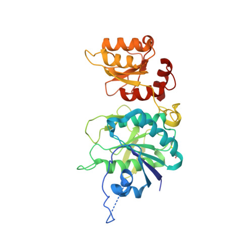
Revealing Escherichia coli type II L-asparaginase active site flexible loop in its open, ligand-free conformation.
Maggi, M., Meli, M., Colombo, G., Scotti, C.(2021) Sci Rep 11: 18885-18885
- PubMed: 34556749
- DOI: https://doi.org/10.1038/s41598-021-98455-1
- Primary Citation of Related Structures:
7P9C - PubMed Abstract:
Since 1993, when the structure of Escherichia coli type II L-asparaginase (EcAII) in complex with L-aspartate was firstly reported, many structures of the wild type and mutated enzyme have been deposited in the Protein Data Bank. None of them report the full structure of the monomer in its ligand-free, open conformation, mainly because of the high dynamic and flexibility of the active site flexible loop. Here we report for the first time the structure of EcAII wild type in its open conformation comprising, for at least one protomer, clear electron density for the active site flexible loop (PDB ID: 6YZI). The structural element is highly mobile and it is transposed onto the rigid part of the active site upon substrate binding to allow completion of the enzyme catalytic center, thanks to key residues that serve as hinges and anchoring points. In the substrate binding pocket, several highly conserved water molecules are coordinated by residues involved in substrate binding, comprising two water molecules very likely involved in the enzyme catalytic process. We also describe, by molecular dynamics simulations, how the transposition of the loop, besides providing the proximity of residues needed for catalysis, causes a general stabilization of the protein.
Organizational Affiliation:
Unit of Immunology and General Pathology, Department of Molecular Medicine, University of Pavia, Via Adolfo Ferrata, 9, 27100, Pavia, Italy.