7P52

GlnK1 from Methanocaldococcus jannaschii with Mg-ATP and 2-oxoglutarate at a resolution of 1.2 A


Experimental Data Snapshot

  • Method: X-RAY DIFFRACTION
  • Resolution: 1.20 Å
  • R-Value Free: 0.155 
  • R-Value Work: 0.131 
  • R-Value Observed: 0.132 

wwPDB Validation   3D Report Full Report


Ligand Structure Quality Assessment 


This is version 1.1 of the entry. See complete history


Literature

The Oxoglutarate Binding Site and Regulatory Mechanism Are Conserved in Ammonium Transporter Inhibitors GlnKs from Methanococcales .

Muller, M.C.Wagner, T.

(2021) Int J Mol Sci 22

  • DOI: https://doi.org/10.3390/ijms22168631
  • Primary Citation of Related Structures:  
    7P4V, 7P4Y, 7P50, 7P52

  • PubMed Abstract: 

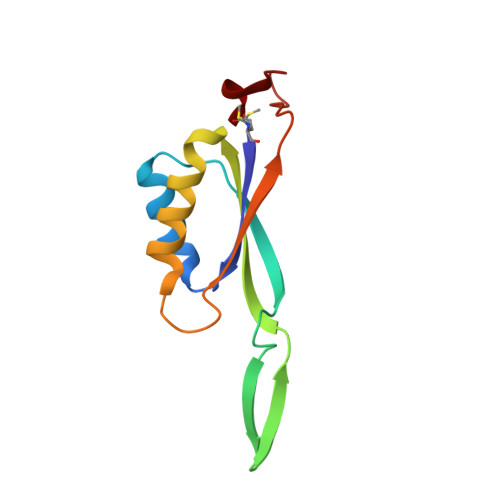
    Protein inhibition is a natural regulatory process to control cellular metabolic fluxes. P II -family signal-transducing effectors are in this matter key regulators of the nitrogen metabolism. Their interaction with their various targets is governed by the cellular nitrogen level and the energy charge. Structural studies on GlnK, a P II -family inhibitor of the ammonium transporters (Amt), showed that the T-loops responsible for channel obstruction are displaced upon the binding of 2-oxoglutarate, magnesium and ATP in a conserved cleft. However, GlnK from Methanocaldococcus jannaschii was shown to bind 2-oxoglutarate on the tip of its T-loop, causing a moderate disruption to GlnK-Amt interaction, raising the question if methanogenic archaea use a singular adaptive strategy. Here we show that membrane fractions of Methanothermococcus thermolithotrophicus released GlnKs only in the presence of Mg-ATP and 2-oxoglutarate. This observation led us to structurally characterize the two GlnK isoforms apo or in complex with ligands. Together, our results show that the 2-oxoglutarate binding interface is conserved in GlnKs from Methanococcales , including Methanocaldococcus jannaschii , emphasizing the importance of a free carboxy-terminal group to facilitate ligand binding and to provoke the shift of the T-loop positions.


  • Organizational Affiliation

    Microbial Metabolism Research Group, Max Planck Institute for Marine Microbiology, Celsiusstraße 1, 28359 Bremen, Germany.


Macromolecules
Find similar proteins by:  (by identity cutoff)  |  3D Structure
Entity ID: 1
MoleculeChains Sequence LengthOrganismDetailsImage
Nitrogen regulatory protein GlnK1112Methanocaldococcus jannaschii DSM 2661Mutation(s): 0 
Gene Names: glnK1MJ0059
UniProt
Find proteins for Q60381 (Methanocaldococcus jannaschii (strain ATCC 43067 / DSM 2661 / JAL-1 / JCM 10045 / NBRC 100440))
Explore Q60381 
Go to UniProtKB:  Q60381
Entity Groups  
Sequence Clusters30% Identity50% Identity70% Identity90% Identity95% Identity100% Identity
UniProt GroupQ60381
Sequence Annotations
Expand
  • Reference Sequence
Small Molecules
Ligands 5 Unique
IDChains Name / Formula / InChI Key2D Diagram3D Interactions
ATP (Subject of Investigation/LOI)
Query on ATP

Download Ideal Coordinates CCD File 
B [auth A]ADENOSINE-5'-TRIPHOSPHATE
C10 H16 N5 O13 P3
ZKHQWZAMYRWXGA-KQYNXXCUSA-N
AKG (Subject of Investigation/LOI)
Query on AKG

Download Ideal Coordinates CCD File 
C [auth A]2-OXOGLUTARIC ACID
C5 H6 O5
KPGXRSRHYNQIFN-UHFFFAOYSA-N
GOL
Query on GOL

Download Ideal Coordinates CCD File 
D [auth A]GLYCEROL
C3 H8 O3
PEDCQBHIVMGVHV-UHFFFAOYSA-N
NO3
Query on NO3

Download Ideal Coordinates CCD File 
E [auth A]NITRATE ION
N O3
NHNBFGGVMKEFGY-UHFFFAOYSA-N
MG (Subject of Investigation/LOI)
Query on MG

Download Ideal Coordinates CCD File 
F [auth A]MAGNESIUM ION
Mg
JLVVSXFLKOJNIY-UHFFFAOYSA-N
Modified Residues  1 Unique
IDChains TypeFormula2D DiagramParent
FME
Query on FME
A
L-PEPTIDE LINKINGC6 H11 N O3 SMET
Experimental Data & Validation

Experimental Data

  • Method: X-RAY DIFFRACTION
  • Resolution: 1.20 Å
  • R-Value Free: 0.155 
  • R-Value Work: 0.131 
  • R-Value Observed: 0.132 
  • Space Group: H 3 2
Unit Cell:
Length ( Å )Angle ( ˚ )
a = 89.053α = 90
b = 89.053β = 90
c = 98.059γ = 120
Software Package:
Software NamePurpose
PHENIXrefinement
PDB_EXTRACTdata extraction
autoPROCdata reduction
autoPROCdata scaling
MOLREPphasing

Structure Validation

View Full Validation Report


Ligand Structure Quality Assessment 


Entry History & Funding Information

Deposition Data


Funding OrganizationLocationGrant Number
Max Planck SocietyGermany--

Revision History  (Full details and data files)

  • Version 1.0: 2021-10-06
    Type: Initial release
  • Version 1.1: 2024-01-31
    Changes: Data collection, Refinement description