7OUJ

Crystal structure of the flavoprotein monooxygenase RubL from rubromycin biosynthesis


Experimental Data Snapshot

  • Method: X-RAY DIFFRACTION
  • Resolution: 1.57 Å
  • R-Value Free: 0.209 
  • R-Value Work: 0.183 

wwPDB Validation   3D Report Full Report


Ligand Structure Quality Assessment 


This is version 1.2 of the entry. See complete history


Literature

Catalytic Control of Spiroketal Formation in Rubromycin Polyketide Biosynthesis.

Toplak, M.Saleem-Batcha, R.Piel, J.Teufel, R.

(2021) Angew Chem Int Ed Engl 60: 26960-26970

  • DOI: https://doi.org/10.1002/anie.202109384
  • Primary Citation of Related Structures:  
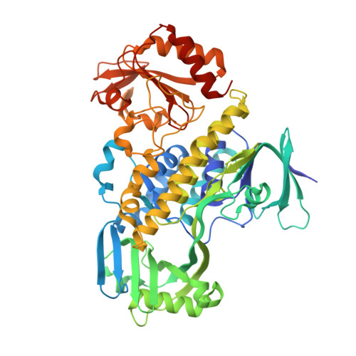
    7OUC, 7OUD, 7OUJ

  • PubMed Abstract: 

    The medically important bacterial aromatic polyketide natural products typically feature a planar, polycyclic core structure. An exception is found for the rubromycins, whose backbones are disrupted by a bisbenzannulated [5,6]-spiroketal pharmacophore that was recently shown to be assembled by flavin-dependent enzymes. In particular, a flavoprotein monooxygenase proved critical for the drastic oxidative rearrangement of a pentangular precursor and the installment of an intermediate [6,6]-spiroketal moiety. Here we provide structural and mechanistic insights into the control of catalysis by this spiroketal synthase, which fulfills several important functions as reductase, monooxygenase, and presumably oxidase. The enzyme hereby tightly controls the redox state of the substrate to counteract shunt product formation, while also steering the cleavage of three carbon-carbon bonds. Our work illustrates an exceptional strategy for the biosynthesis of stable chroman spiroketals.


  • Organizational Affiliation

    Faculty of Biology, University of Freiburg, Schänzlestrasse 1, 79104, Freiburg, Germany.


Macromolecules
Find similar proteins by:  (by identity cutoff)  |  3D Structure
Entity ID: 1
MoleculeChains Sequence LengthOrganismDetailsImage
RubLA [auth AAA]555Streptomyces collinusMutation(s): 0 
Gene Names: rubL
UniProt
Find proteins for Q8KY42 (Streptomyces collinus)
Explore Q8KY42 
Go to UniProtKB:  Q8KY42
Entity Groups  
Sequence Clusters30% Identity50% Identity70% Identity90% Identity95% Identity100% Identity
UniProt GroupQ8KY42
Sequence Annotations
Expand
  • Reference Sequence
Small Molecules
Ligands 5 Unique
IDChains Name / Formula / InChI Key2D Diagram3D Interactions
FAD (Subject of Investigation/LOI)
Query on FAD

Download Ideal Coordinates CCD File 
B [auth AAA]FLAVIN-ADENINE DINUCLEOTIDE
C27 H33 N9 O15 P2
VWWQXMAJTJZDQX-UYBVJOGSSA-N
1JW
Query on 1JW

Download Ideal Coordinates CCD File 
D [auth AAA],
F [auth AAA],
G [auth AAA]
(2S)-hexane-1,2,6-triol
C6 H14 O3
ZWVMLYRJXORSEP-LURJTMIESA-N
HYP
Query on HYP

Download Ideal Coordinates CCD File 
E [auth AAA]4-HYDROXYPROLINE
C5 H9 N O3
PMMYEEVYMWASQN-DMTCNVIQSA-N
GOL
Query on GOL

Download Ideal Coordinates CCD File 
H [auth AAA]GLYCEROL
C3 H8 O3
PEDCQBHIVMGVHV-UHFFFAOYSA-N
CL (Subject of Investigation/LOI)
Query on CL

Download Ideal Coordinates CCD File 
C [auth AAA]CHLORIDE ION
Cl
VEXZGXHMUGYJMC-UHFFFAOYSA-M
Experimental Data & Validation

Experimental Data

  • Method: X-RAY DIFFRACTION
  • Resolution: 1.57 Å
  • R-Value Free: 0.209 
  • R-Value Work: 0.183 
  • Space Group: I 1 2 1
Unit Cell:
Length ( Å )Angle ( ˚ )
a = 77.323α = 90
b = 57.514β = 95.183
c = 122.379γ = 90
Software Package:
Software NamePurpose
REFMACrefinement
XDSdata reduction
SCALAdata scaling
PHASERphasing

Structure Validation

View Full Validation Report


Ligand Structure Quality Assessment 


Entry History & Funding Information

Deposition Data


Funding OrganizationLocationGrant Number
German Research Foundation (DFG)GermanyTE 931/3-1
German Research Foundation (DFG)GermanyTE 931/4-1
Austrian Science FundAustriaJ4482-B

Revision History  (Full details and data files)

  • Version 1.0: 2021-11-03
    Type: Initial release
  • Version 1.1: 2021-12-22
    Changes: Database references
  • Version 1.2: 2024-01-31
    Changes: Data collection, Derived calculations, Refinement description