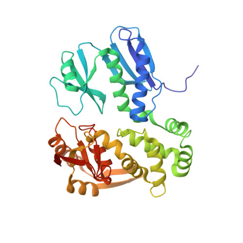
Structural Dynamics of the Functional Nonameric Type III Translocase Export Gate.
Yuan, B., Portaliou, A.G., Parakra, R., Smit, J.H., Wald, J., Li, Y., Srinivasu, B., Loos, M.S., Dhupar, H.S., Fahrenkamp, D., Kalodimos, C.G., Duong van Hoa, F., Cordes, T., Karamanou, S., Marlovits, T.C., Economou, A.(2021) J Mol Biol 433: 167188-167188
- PubMed: 34454944
- DOI: https://doi.org/10.1016/j.jmb.2021.167188
- Primary Citation of Related Structures:
7OSL - PubMed Abstract:
Type III protein secretion is widespread in Gram-negative pathogens. It comprises the injectisome with a surface-exposed needle and an inner membrane translocase. The translocase contains the SctRSTU export channel enveloped by the export gate subunit SctV that binds chaperone/exported clients and forms a putative ante-chamber. We probed the assembly, function, structure and dynamics of SctV from enteropathogenic E. coli (EPEC). In both EPEC and E. coli lab strains, SctV forms peripheral oligomeric clusters that are detergent-extracted as homo-nonamers. Membrane-embedded SctV 9 is necessary and sufficient to act as a receptor for different chaperone/exported protein pairs with distinct C-domain binding sites that are essential for secretion. Negative staining electron microscopy revealed that peptidisc-reconstituted His-SctV 9 forms a tripartite particle of ∼22 nm with a N-terminal domain connected by a short linker to a C-domain ring structure with a ∼5 nm-wide inner opening. The isolated C-domain ring was resolved with cryo-EM at 3.1 Å and structurally compared to other SctV homologues. Its four sub-domains undergo a three-stage "pinching" motion. Hydrogen-deuterium exchange mass spectrometry revealed this to involve dynamic and rigid hinges and a hyper-flexible sub-domain that flips out of the ring periphery and binds chaperones on and between adjacent protomers. These motions are coincident with local conformational changes at the pore surface and ring entry mouth that may also be modulated by the ATPase inner stalk. We propose that the intrinsic dynamics of the SctV protomer are modulated by chaperones and the ATPase and could affect allosterically the other subunits of the nonameric ring during secretion.
Organizational Affiliation:
KU Leuven, Department of Microbiology and Immunology, Rega Institute for Medical Research, Laboratory of Molecular Bacteriology, B-3000 Leuven, Belgium; Centre for Structural Systems Biology (CSSB), Notkestrasse 85, D-22607 Hamburg, Germany; University Medical Center Hamburg-Eppendorf (UKE), Institute for Structural and Systems Biology, Notkestrasse 85, D-22607 Hamburg, Germany; German Electron Synchrotron Centre (DESY), Notkestrasse 85, D-22607 Hamburg, Germany. Electronic address: biao.yuan@cssb-hamburg.de.


