7OMT

Crystal structure of ProMacrobody 21 with bound maltose


Experimental Data Snapshot

  • Method: X-RAY DIFFRACTION
  • Resolution: 2.00 Å
  • R-Value Free: 0.239 
  • R-Value Work: 0.193 
  • R-Value Observed: 0.195 

wwPDB Validation   3D Report Full Report


This is version 1.2 of the entry. See complete history


Literature

Cryo-EM structures of a LptDE transporter in complex with Pro-macrobodies offer insight into lipopolysaccharide translocation.

Botte, M.Ni, D.Schenck, S.Zimmermann, I.Chami, M.Bocquet, N.Egloff, P.Bucher, D.Trabuco, M.Cheng, R.K.Y.Brunner, J.D.Seeger, M.A.Stahlberg, H.Hennig, M.

(2022) Nat Commun 13: 1826-1826

  • DOI: https://doi.org/10.1038/s41467-022-29459-2
  • Primary Citation of Related Structures:  
    7OMM, 7OMT

  • PubMed Abstract: 

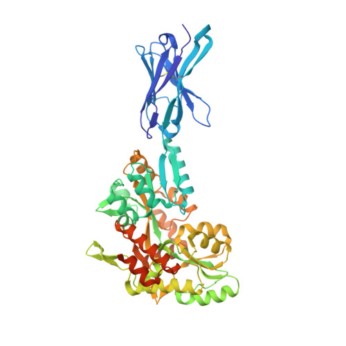
    Lipopolysaccharides are major constituents of the extracellular leaflet in the bacterial outer membrane and form an effective physical barrier for environmental threats and for antibiotics in Gram-negative bacteria. The last step of LPS insertion via the Lpt pathway is mediated by the LptD/E protein complex. Detailed insights into the architecture of LptDE transporter complexes have been derived from X-ray crystallography. However, no structure of a laterally open LptD transporter, a transient state that occurs during LPS release, is available to date. Here, we report a cryo-EM structure of a partially opened LptDE transporter in complex with rigid chaperones derived from nanobodies, at 3.4 Å resolution. In addition, a subset of particles allows to model a structure of a laterally fully opened LptDE complex. Our work offers insights into the mechanism of LPS insertion, provides a structural framework for the development of antibiotics targeting LptD and describes a highly rigid chaperone scaffold to enable structural biology of challenging protein targets.


  • Organizational Affiliation

    leadXpro AG, Park Innovaare, 5234, Villigen, Switzerland.


Macromolecules
Find similar proteins by:  (by identity cutoff)  |  3D Structure
Entity ID: 1
MoleculeChains Sequence LengthOrganismDetailsImage
ProMacrobody 21520synthetic constructMutation(s): 0 
Membrane Entity: Yes 
UniProt
Find proteins for P0AEX9 (Escherichia coli (strain K12))
Explore P0AEX9 
Go to UniProtKB:  P0AEX9
Entity Groups  
Sequence Clusters30% Identity50% Identity70% Identity90% Identity95% Identity100% Identity
UniProt GroupP0AEX9
Sequence Annotations
Expand
  • Reference Sequence
Oligosaccharides

Help

Entity ID: 2
MoleculeChains Length2D Diagram Glycosylation3D Interactions
alpha-D-glucopyranose-(1-4)-alpha-D-glucopyranose
B
2N/A
Glycosylation Resources
GlyTouCan:  G07411ON
GlyCosmos:  G07411ON
Biologically Interesting Molecules (External Reference) 1 Unique
Experimental Data & Validation

Experimental Data

  • Method: X-RAY DIFFRACTION
  • Resolution: 2.00 Å
  • R-Value Free: 0.239 
  • R-Value Work: 0.193 
  • R-Value Observed: 0.195 
  • Space Group: I 1 2 1
Unit Cell:
Length ( Å )Angle ( ˚ )
a = 51.897α = 90
b = 87.624β = 99.076
c = 118.635γ = 90
Software Package:
Software NamePurpose
REFMACrefinement
PHENIXrefinement
XDSdata reduction
PHASERphasing

Structure Validation

View Full Validation Report


Entry History & Funding Information

Deposition Data


Funding OrganizationLocationGrant Number
InnosuisseSwitzerland25864.1 PFLS-LS

Revision History  (Full details and data files)

  • Version 1.0: 2022-05-04
    Type: Initial release
  • Version 1.1: 2022-05-11
    Changes: Database references
  • Version 1.2: 2024-01-31
    Changes: Data collection, Refinement description