7OM8

Beta2 appendage domain of AP2 bound to terminal domains beneath the hub of the 28 triskelia mini clathrin coat complex, class 15


Experimental Data Snapshot

  • Method: ELECTRON MICROSCOPY
  • Resolution: 10.5 Å
  • Aggregation State: PARTICLE 
  • Reconstruction Method: SINGLE PARTICLE 

wwPDB Validation   3D Report Full Report


This is version 3.0 of the entry. See complete history


Literature

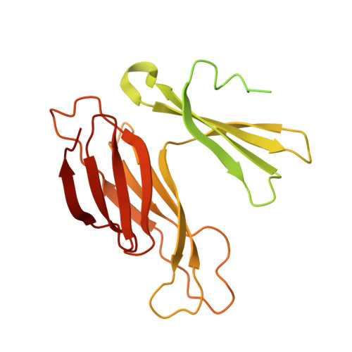
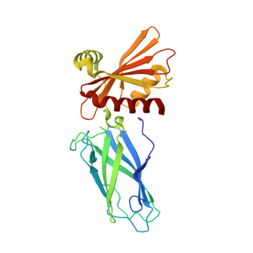
Multi-modal adaptor-clathrin contacts drive coated vesicle assembly.

Smith, S.M.Larocque, G.Wood, K.M.Morris, K.L.Roseman, A.M.Sessions, R.B.Royle, S.J.Smith, C.J.

(2021) EMBO J 40: e108795-e108795

  • DOI: https://doi.org/10.15252/embj.2021108795
  • Primary Citation of Related Structures:  
    7OM8

  • PubMed Abstract: 

    Clathrin-coated pits are formed by the recognition of membrane and cargo by the AP2 complex and the subsequent recruitment of clathrin triskelia. A role for AP2 in coated-pit assembly beyond initial clathrin recruitment has not been explored. Clathrin binds the β2 subunit of AP2, and several binding sites have been identified, but our structural knowledge of these interactions is incomplete and their functional importance during endocytosis is unclear. Here, we analysed the cryo-EM structure of clathrin cages assembled in the presence of β2 hinge-appendage (β2HA). We find that the β2-appendage binds in at least two positions in the cage, demonstrating that multi-modal binding is a fundamental property of clathrin-AP2 interactions. In one position, β2-appendage cross-links two adjacent terminal domains from different triskelia. Functional analysis of β2HA-clathrin interactions reveals that endocytosis requires two clathrin interaction sites: a clathrin-box motif on the hinge and the "sandwich site" on the appendage. We propose that β2-appendage binding to more than one triskelion is a key feature of the system and likely explains why assembly is driven by AP2.


  • Organizational Affiliation

    School of Life Sciences, University of Warwick, Coventry, UK.


Macromolecules
Find similar proteins by:  (by identity cutoff)  |  3D Structure
Entity ID: 1
MoleculeChains Sequence LengthOrganismDetailsImage
Clathrin heavy chainA [auth Y],
B [auth Z]
299Sus scrofaMutation(s): 0 
UniProt
Find proteins for I3LGD4 (Sus scrofa)
Explore I3LGD4 
Go to UniProtKB:  I3LGD4
Entity Groups  
Sequence Clusters30% Identity50% Identity70% Identity90% Identity95% Identity100% Identity
UniProt GroupI3LGD4
Sequence Annotations
Expand
  • Reference Sequence
Find similar proteins by:  (by identity cutoff)  |  3D Structure
Entity ID: 2
MoleculeChains Sequence LengthOrganismDetailsImage
AP-2 complex subunit betaC [auth B]233Homo sapiensMutation(s): 0 
Gene Names: AP2B1ADTB2CLAPB1
UniProt & NIH Common Fund Data Resources
Find proteins for P63010 (Homo sapiens)
Explore P63010 
Go to UniProtKB:  P63010
PHAROS:  P63010
GTEx:  ENSG00000006125 
Entity Groups  
Sequence Clusters30% Identity50% Identity70% Identity90% Identity95% Identity100% Identity
UniProt GroupP63010
Sequence Annotations
Expand
  • Reference Sequence
Experimental Data & Validation

Experimental Data

  • Method: ELECTRON MICROSCOPY
  • Resolution: 10.5 Å
  • Aggregation State: PARTICLE 
  • Reconstruction Method: SINGLE PARTICLE 

Structure Validation

View Full Validation Report


Entry History & Funding Information

Deposition Data


Funding OrganizationLocationGrant Number
Biotechnology and Biological Sciences Research Council (BBSRC)United KingdomBB/N008391/1

Revision History  (Full details and data files)

  • Version 1.0: 2021-08-11
    Type: Initial release
  • Version 1.1: 2021-09-29
    Changes: Data collection, Database references
  • Version 2.0: 2021-11-17
    Type: Coordinate replacement
    Reason: Atomic clashes
    Changes: Advisory, Atomic model, Data collection, Database references, Derived calculations, Other, Refinement description, Structure summary
  • Version 3.0: 2022-07-13
    Type: Coordinate replacement
    Reason: Chirality error
    Changes: Advisory, Atomic model, Data collection, Source and taxonomy