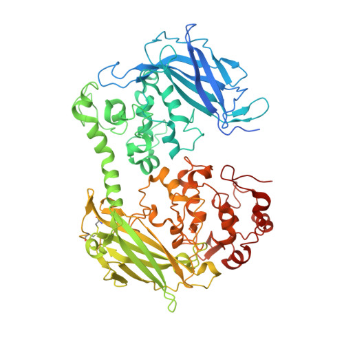
7O7G

Crystal structure of the Shewanella oneidensis MR1 MtrC mutant H561M


Experimental Data Snapshot

  • Method: X-RAY DIFFRACTION
  • Resolution: 1.60 Å
  • R-Value Free: 0.205 
  • R-Value Work: 0.170 
  • R-Value Observed: 0.172 

wwPDB Validation   3D Report Full Report


Ligand Structure Quality Assessment 


This is version 1.1 of the entry. See complete history


Literature

Nanosecond heme-to-heme electron transfer rates in a multiheme cytochrome nanowire reported by a spectrally unique His/Met-ligated heme.

van Wonderen, J.H.Adamczyk, K.Wu, X.Jiang, X.Piper, S.E.H.Hall, C.R.Edwards, M.J.Clarke, T.A.Zhang, H.Jeuken, L.J.C.Sazanovich, I.V.Towrie, M.Blumberger, J.Meech, S.R.Butt, J.N.

(2021) Proc Natl Acad Sci U S A 118

  • DOI: https://doi.org/10.1073/pnas.2107939118
  • Primary Citation of Related Structures:  
    7O7G

  • PubMed Abstract: 

    Proteins achieve efficient energy storage and conversion through electron transfer along a series of redox cofactors. Multiheme cytochromes are notable examples. These proteins transfer electrons over distance scales of several nanometers to >10 μm and in so doing they couple cellular metabolism with extracellular redox partners including electrodes. Here, we report pump-probe spectroscopy that provides a direct measure of the intrinsic rates of heme-heme electron transfer in this fascinating class of proteins. Our study took advantage of a spectrally unique His/Met-ligated heme introduced at a defined site within the decaheme extracellular MtrC protein of Shewanella oneidensis We observed rates of heme-to-heme electron transfer on the order of 10 9 s -1 (3.7 to 4.3 Å edge-to-edge distance), in good agreement with predictions based on density functional and molecular dynamics calculations. These rates are among the highest reported for ground-state electron transfer in biology. Yet, some fall 2 to 3 orders of magnitude below the Moser-Dutton ruler because electron transfer at these short distances is through space and therefore associated with a higher tunneling barrier than the through-protein tunneling scenario that is usual at longer distances. Moreover, we show that the His/Met-ligated heme creates an electron sink that stabilizes the charge separated state on the 100-μs time scale. This feature could be exploited in future designs of multiheme cytochromes as components of versatile photosynthetic biohybrid assemblies.


  • Organizational Affiliation

    School of Chemistry, University of East Anglia, Norwich Research Park, Norwich NR4 7TJ, United Kingdom.


Macromolecules
Find similar proteins by:  (by identity cutoff)  |  3D Structure
Entity ID: 1
MoleculeChains Sequence LengthOrganismDetailsImage
Extracellular iron oxide respiratory system surface decaheme cytochrome c component MtrC671Shewanella oneidensis MR-1Mutation(s): 1 
Gene Names: mtrCSO_1778
UniProt
Find proteins for Q8EG34 (Shewanella oneidensis (strain MR-1))
Explore Q8EG34 
Go to UniProtKB:  Q8EG34
Entity Groups  
Sequence Clusters30% Identity50% Identity70% Identity90% Identity95% Identity100% Identity
UniProt GroupQ8EG34
Sequence Annotations
Expand
  • Reference Sequence
Small Molecules
Ligands 4 Unique
IDChains Name / Formula / InChI Key2D Diagram3D Interactions
HEC
Query on HEC

Download Ideal Coordinates CCD File 
B [auth A]
C [auth A]
D [auth A]
E [auth A]
F [auth A]
B [auth A],
C [auth A],
D [auth A],
E [auth A],
F [auth A],
G [auth A],
H [auth A],
I [auth A],
J [auth A],
K [auth A]
HEME C
C34 H34 Fe N4 O4
HXQIYSLZKNYNMH-LJNAALQVSA-N
EDO
Query on EDO

Download Ideal Coordinates CCD File 
BA [auth A]
P [auth A]
Q [auth A]
R [auth A]
S [auth A]
BA [auth A],
P [auth A],
Q [auth A],
R [auth A],
S [auth A],
T [auth A],
U [auth A],
V [auth A]
1,2-ETHANEDIOL
C2 H6 O2
LYCAIKOWRPUZTN-UHFFFAOYSA-N
ACT
Query on ACT

Download Ideal Coordinates CCD File 
AA [auth A],
W [auth A],
X [auth A],
Y [auth A],
Z [auth A]
ACETATE ION
C2 H3 O2
QTBSBXVTEAMEQO-UHFFFAOYSA-M
CA
Query on CA

Download Ideal Coordinates CCD File 
L [auth A],
M [auth A],
N [auth A],
O [auth A]
CALCIUM ION
Ca
BHPQYMZQTOCNFJ-UHFFFAOYSA-N
Experimental Data & Validation

Experimental Data

  • Method: X-RAY DIFFRACTION
  • Resolution: 1.60 Å
  • R-Value Free: 0.205 
  • R-Value Work: 0.170 
  • R-Value Observed: 0.172 
  • Space Group: P 21 21 21
Unit Cell:
Length ( Å )Angle ( ˚ )
a = 52.875α = 90
b = 89.657β = 90
c = 153.9γ = 90
Software Package:
Software NamePurpose
REFMACrefinement
PDB_EXTRACTdata extraction
DIALSdata reduction
DIALSdata scaling
PHASERphasing

Structure Validation

View Full Validation Report


Ligand Structure Quality Assessment 


Entry History & Funding Information

Deposition Data


Funding OrganizationLocationGrant Number
Biotechnology and Biological Sciences Research Council (BBSRC)United KingdomBB/P01819X/1
Biotechnology and Biological Sciences Research Council (BBSRC)United KingdomBB/S002499/1
Engineering and Physical Sciences Research CouncilUnited KingdomEP/M001989/1

Revision History  (Full details and data files)

  • Version 1.0: 2021-10-06
    Type: Initial release
  • Version 1.1: 2024-01-31
    Changes: Data collection, Refinement description