7O50

Crystal structure of human legumain in complex with Gly-Ser-Asn peptide


Experimental Data Snapshot

  • Method: X-RAY DIFFRACTION
  • Resolution: 1.90 Å
  • R-Value Free: 0.219 
  • R-Value Work: 0.192 
  • R-Value Observed: 0.194 

wwPDB Validation   3D Report Full Report


Ligand Structure Quality Assessment 


This is version 2.1 of the entry. See complete history


Literature

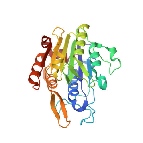
The Peptide Ligase Activity of Human Legumain Depends on Fold Stabilization and Balanced Substrate Affinities.

Dall, E.Stanojlovic, V.Demir, F.Briza, P.Dahms, S.O.Huesgen, P.F.Cabrele, C.Brandstetter, H.

(2021) ACS Catal 11: 11885-11896

  • DOI: https://doi.org/10.1021/acscatal.1c02057
  • Primary Citation of Related Structures:  
    7O50

  • PubMed Abstract: 

    Protein modification by enzymatic breaking and forming of peptide bonds significantly expands the repertoire of genetically encoded protein sequences. The dual protease-ligase legumain exerts the two opposing activities within a single protein scaffold. Primarily localized to the endolysosomal system, legumain represents a key enzyme in the generation of antigenic peptides for subsequent presentation on the MHCII complex. Here we show that human legumain catalyzes the ligation and cyclization of linear peptides at near-neutral pH conditions, where legumain is intrinsically unstable. Conformational stabilization significantly enhanced legumain's ligase activity, which further benefited from engineering the prime substrate recognition sites for improved affinity. Additionally, we provide evidence that specific legumain activation states allow for differential regulation of its activities. Together these results set the basis for engineering legumain proteases and ligases with applications in biotechnology and drug development.


  • Organizational Affiliation

    Department of Biosciences, University of Salzburg, 5020 Salzburg, Austria.


Macromolecules
Find similar proteins by:  (by identity cutoff)  |  3D Structure
Entity ID: 1
MoleculeChains Sequence LengthOrganismDetailsImage
Legumain
A, B
262Homo sapiensMutation(s): 1 
Gene Names: LGMNPRSC1
EC: 3.4.22.34
UniProt & NIH Common Fund Data Resources
Find proteins for Q99538 (Homo sapiens)
Explore Q99538 
Go to UniProtKB:  Q99538
PHAROS:  Q99538
GTEx:  ENSG00000100600 
Entity Groups  
Sequence Clusters30% Identity50% Identity70% Identity90% Identity95% Identity100% Identity
UniProt GroupQ99538
Sequence Annotations
Expand
  • Reference Sequence

Find similar proteins by:  Sequence   |   3D Structure  

Entity ID: 2
MoleculeChains Sequence LengthOrganismDetailsImage
GLY-SER-ASNC [auth H],
D [auth C]
4synthetic constructMutation(s): 0 
Sequence Annotations
Expand
  • Reference Sequence
Oligosaccharides

Help

Entity ID: 3
MoleculeChains Length2D Diagram Glycosylation3D Interactions
2-acetamido-2-deoxy-beta-D-glucopyranose-(1-4)-2-acetamido-2-deoxy-beta-D-glucopyranoseE [auth G],
F [auth J],
G [auth P]
2N-Glycosylation
Glycosylation Resources
GlyTouCan:  G42666HT
GlyCosmos:  G42666HT
GlyGen:  G42666HT
Small Molecules
Ligands 1 Unique
IDChains Name / Formula / InChI Key2D Diagram3D Interactions
NAG
Query on NAG

Download Ideal Coordinates CCD File 
H [auth A],
I [auth B]
2-acetamido-2-deoxy-beta-D-glucopyranose
C8 H15 N O6
OVRNDRQMDRJTHS-FMDGEEDCSA-N
Modified Residues  1 Unique
IDChains TypeFormula2D DiagramParent
SNN
Query on SNN
A, B
L-PEPTIDE LINKINGC4 H6 N2 O2ASN
Experimental Data & Validation

Experimental Data

  • Method: X-RAY DIFFRACTION
  • Resolution: 1.90 Å
  • R-Value Free: 0.219 
  • R-Value Work: 0.192 
  • R-Value Observed: 0.194 
  • Space Group: P 21 21 21
Unit Cell:
Length ( Å )Angle ( ˚ )
a = 43.36α = 90
b = 75.11β = 90
c = 172.31γ = 90
Software Package:
Software NamePurpose
PHENIXrefinement
PHENIXrefinement
iMOSFLMdata reduction
Aimlessdata scaling
PHENIXphasing

Structure Validation

View Full Validation Report


Ligand Structure Quality Assessment 


Entry History & Funding Information

Deposition Data


Funding OrganizationLocationGrant Number
Austrian Science FundAustriaP31867
Austrian Science FundAustriaW_01213

Revision History  (Full details and data files)

  • Version 1.0: 2021-09-22
    Type: Initial release
  • Version 1.1: 2021-10-20
    Changes: Data collection, Database references
  • Version 2.0: 2023-11-15
    Changes: Atomic model, Data collection, Derived calculations
  • Version 2.1: 2024-01-31
    Changes: Refinement description