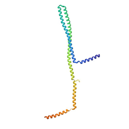
7O40

Structural basis for VIPP1 oligomerization and maintenance of thylakoid membrane integrity


Experimental Data Snapshot

  • Method: ELECTRON MICROSCOPY
  • Resolution: 4.30 Å
  • Aggregation State: PARTICLE 
  • Reconstruction Method: SINGLE PARTICLE 

wwPDB Validation   3D Report Full Report


This is version 1.2 of the entry. See complete history


Literature

Structural basis for VIPP1 oligomerization and maintenance of thylakoid membrane integrity.

Gupta, T.K.Klumpe, S.Gries, K.Heinz, S.Wietrzynski, W.Ohnishi, N.Niemeyer, J.Spaniol, B.Schaffer, M.Rast, A.Ostermeier, M.Strauss, M.Plitzko, J.M.Baumeister, W.Rudack, T.Sakamoto, W.Nickelsen, J.Schuller, J.M.Schroda, M.Engel, B.D.

(2021) Cell 184: 3643

  • DOI: https://doi.org/10.1016/j.cell.2021.05.011
  • Primary Citation of Related Structures:  
    7O3W, 7O3X, 7O3Y, 7O3Z, 7O40

  • PubMed Abstract: 

    Vesicle-inducing protein in plastids 1 (VIPP1) is essential for the biogenesis and maintenance of thylakoid membranes, which transform light into life. However, it is unknown how VIPP1 performs its vital membrane-remodeling functions. Here, we use cryo-electron microscopy to determine structures of cyanobacterial VIPP1 rings, revealing how VIPP1 monomers flex and interweave to form basket-like assemblies of different symmetries. Three VIPP1 monomers together coordinate a non-canonical nucleotide binding pocket on one end of the ring. Inside the ring's lumen, amphipathic helices from each monomer align to form large hydrophobic columns, enabling VIPP1 to bind and curve membranes. In vivo mutations in these hydrophobic surfaces cause extreme thylakoid swelling under high light, indicating an essential role of VIPP1 lipid binding in resisting stress-induced damage. Using cryo-correlative light and electron microscopy (cryo-CLEM), we observe oligomeric VIPP1 coats encapsulating membrane tubules within the Chlamydomonas chloroplast. Our work provides a structural foundation for understanding how VIPP1 directs thylakoid biogenesis and maintenance.


  • Organizational Affiliation

    Department of Molecular Structural Biology, Max Planck Institute of Biochemistry, 82152 Martinsried, Germany.


Macromolecules
Find similar proteins by:  (by identity cutoff)  |  3D Structure
Entity ID: 1
MoleculeChains Sequence LengthOrganismDetailsImage
Protein sll0617266Synechocystis sp. PCC 6803 substr. KazusaMutation(s): 0 
Gene Names: sll0617
UniProt
Find proteins for Q55707 (Synechocystis sp. (strain PCC 6803 / Kazusa))
Explore Q55707 
Go to UniProtKB:  Q55707
Entity Groups  
Sequence Clusters30% Identity50% Identity70% Identity90% Identity95% Identity100% Identity
UniProt GroupQ55707
Sequence Annotations
Expand
  • Reference Sequence
Small Molecules
Ligands 1 Unique
IDChains Name / Formula / InChI Key2D Diagram3D Interactions
ADP (Subject of Investigation/LOI)
Query on ADP

Download Ideal Coordinates CCD File 
G [auth A]ADENOSINE-5'-DIPHOSPHATE
C10 H15 N5 O10 P2
XTWYTFMLZFPYCI-KQYNXXCUSA-N
Experimental Data & Validation

Experimental Data

  • Method: ELECTRON MICROSCOPY
  • Resolution: 4.30 Å
  • Aggregation State: PARTICLE 
  • Reconstruction Method: SINGLE PARTICLE 
EM Software:
TaskSoftware PackageVersion
MODEL REFINEMENTPHENIX1.17.1
RECONSTRUCTIONRELION3.0

Structure Validation

View Full Validation Report


Entry History & Funding Information

Deposition Data


Funding OrganizationLocationGrant Number
German Research Foundation (DFG)GermanyFOR2092 EN1194/1-1
German Research Foundation (DFG)GermanyNI 390/9-2
German Research Foundation (DFG)GermanySCHU 3364
German Research Foundation (DFG)GermanyFOR2092 Schr 617/8-2
German Research Foundation (DFG)GermanyTRR 175/C02
Ministry of Education, Culture, Sports, Science and Technology (Japan)Japan16H06554
Japan Society for the Promotion of Science (JSPS)Japan17H03699

Revision History  (Full details and data files)

  • Version 1.0: 2021-06-30
    Type: Initial release
  • Version 1.1: 2021-07-07
    Changes: Database references
  • Version 1.2: 2021-07-21
    Changes: Database references