7O22

X-ray structure of furin in complex with the guanylhydrazone-based inhibitor 4 (mi359)


Experimental Data Snapshot

  • Method: X-RAY DIFFRACTION
  • Resolution: 1.80 Å
  • R-Value Free: 0.182 
  • R-Value Work: 0.169 
  • R-Value Observed: 0.170 

wwPDB Validation   3D Report Full Report


This is version 1.2 of the entry. See complete history


Literature

OFF-State-Specific Inhibition of the Proprotein Convertase Furin.

Dahms, S.O.Haider, T.Klebe, G.Steinmetzer, T.Brandstetter, H.

(2021) ACS Chem Biol 16: 1692-1700

  • DOI: https://doi.org/10.1021/acschembio.1c00411
  • Primary Citation of Related Structures:  
    7O1U, 7O1W, 7O1Y, 7O20, 7O22

  • PubMed Abstract: 

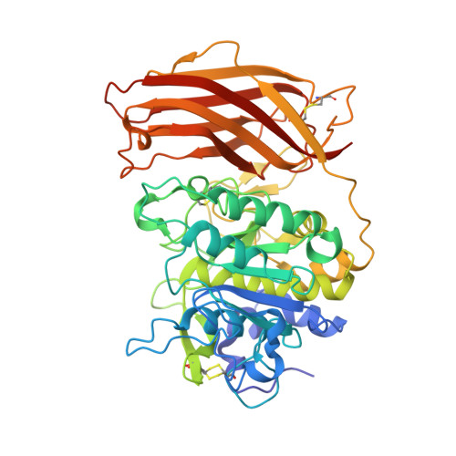
    The pro-protein convertase furin is a highly specific serine protease involved in the proteolytic maturation of many proteins in the secretory pathway. It also activates surface proteins of many viruses including the severe acute respiratory syndrome coronavirus 2 (SARS-CoV-2). Furin inhibitors effectively suppress viral replication and thus are promising antiviral therapeutics with broad application potential. Polybasic substrate-like ligands typically trigger conformational changes shifting furin's active site cleft from the OFF-state to the ON-state. Here, we solved the X-ray structures of furin in complex with four different arginine mimetic compounds with reduced basicity. These guanylhydrazone-based inhibitor complexes showed for the first time an active site-directed binding mode to furin's OFF-state conformation. The compounds undergo unique interactions within the S1 pocket, largely different compared to substrate-like ligands. A second binding site was identified at the S4/S5 pocket of furin. Crystallography-based titration experiments confirmed the S1 site as the primary binding pocket. We also tested the proprotein convertases PC5/6 and PC7 for inhibition by guanylhydrazones and found an up to 7-fold lower potency for PC7. Interestingly, the observed differences in the K i values correlated with the sequence conservation of the PCs at the allosteric sodium binding site. Therefore, OFF-state-specific targeting of furin can serve as a valuable strategy for structure-based development of PC-selective small-molecule inhibitors.


  • Organizational Affiliation

    Department of Biosciences, University of Salzburg, Hellbrunnerstraße 34, A-5020 Salzburg, Austria.


Macromolecules
Find similar proteins by:  (by identity cutoff)  |  3D Structure
Entity ID: 1
MoleculeChains Sequence LengthOrganismDetailsImage
Furin480Homo sapiensMutation(s): 0 
Gene Names: FURINFURPACEPCSK3
EC: 3.4.21.75
UniProt & NIH Common Fund Data Resources
Find proteins for P09958 (Homo sapiens)
Explore P09958 
Go to UniProtKB:  P09958
PHAROS:  P09958
GTEx:  ENSG00000140564 
Entity Groups  
Sequence Clusters30% Identity50% Identity70% Identity90% Identity95% Identity100% Identity
UniProt GroupP09958
Sequence Annotations
Expand
  • Reference Sequence
Small Molecules
Ligands 6 Unique
IDChains Name / Formula / InChI Key2D Diagram3D Interactions
UYW (Subject of Investigation/LOI)
Query on UYW

Download Ideal Coordinates CCD File 
M [auth A][azanyl-[(2E)-2-[[2-[(4-chlorophenyl)methoxy]phenyl]methylidene]hydrazinyl]methylidene]azanium
C15 H16 Cl N4 O
QLZIJPLTRJOVDN-DJKKODMXSA-O
NAG
Query on NAG

Download Ideal Coordinates CCD File 
L [auth A]2-acetamido-2-deoxy-beta-D-glucopyranose
C8 H15 N O6
OVRNDRQMDRJTHS-FMDGEEDCSA-N
DMS
Query on DMS

Download Ideal Coordinates CCD File 
H [auth A],
I [auth A],
J [auth A],
K [auth A]
DIMETHYL SULFOXIDE
C2 H6 O S
IAZDPXIOMUYVGZ-UHFFFAOYSA-N
CA
Query on CA

Download Ideal Coordinates CCD File 
B [auth A],
C [auth A],
D [auth A]
CALCIUM ION
Ca
BHPQYMZQTOCNFJ-UHFFFAOYSA-N
CL
Query on CL

Download Ideal Coordinates CCD File 
G [auth A]CHLORIDE ION
Cl
VEXZGXHMUGYJMC-UHFFFAOYSA-M
NA
Query on NA

Download Ideal Coordinates CCD File 
E [auth A],
F [auth A]
SODIUM ION
Na
FKNQFGJONOIPTF-UHFFFAOYSA-N
Experimental Data & Validation

Experimental Data

  • Method: X-RAY DIFFRACTION
  • Resolution: 1.80 Å
  • R-Value Free: 0.182 
  • R-Value Work: 0.169 
  • R-Value Observed: 0.170 
  • Space Group: P 65 2 2
Unit Cell:
Length ( Å )Angle ( ˚ )
a = 130.976α = 90
b = 130.976β = 90
c = 155.485γ = 120
Software Package:
Software NamePurpose
PHENIXrefinement
PHENIXphasing
XDSdata reduction
XDSdata scaling

Structure Validation

View Full Validation Report


Entry History & Funding Information

Deposition Data


Funding OrganizationLocationGrant Number
Austrian Science FundAustriaM 2730

Revision History  (Full details and data files)

  • Version 1.0: 2021-09-01
    Type: Initial release
  • Version 1.1: 2021-09-29
    Changes: Data collection, Database references
  • Version 1.2: 2024-01-31
    Changes: Data collection, Refinement description