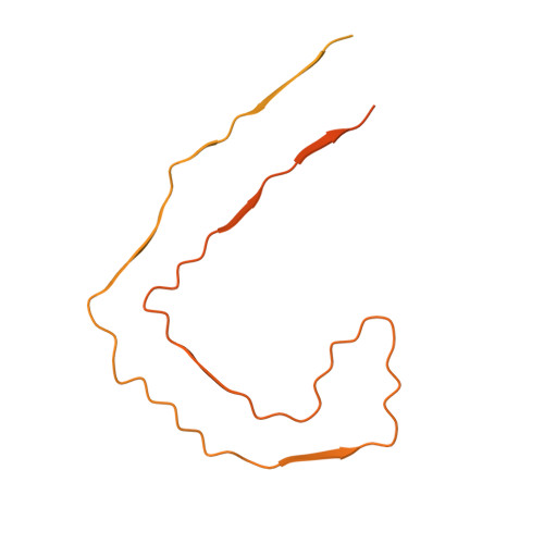
7NRX

Straight filament from Alzheimer's disease with PET ligand APN-1607


Experimental Data Snapshot

  • Method: ELECTRON MICROSCOPY
  • Resolution: 3.55 Å
  • Aggregation State: FILAMENT 
  • Reconstruction Method: HELICAL 

wwPDB Validation   3D Report Full Report


This is version 1.2 of the entry. See complete history


Literature

Cryo-EM structures of tau filaments from Alzheimer's disease with PET ligand APN-1607.

Shi, Y.Murzin, A.G.Falcon, B.Epstein, A.Machin, J.Tempest, P.Newell, K.L.Vidal, R.Garringer, H.J.Sahara, N.Higuchi, M.Ghetti, B.Jang, M.K.Scheres, S.H.W.Goedert, M.

(2021) Acta Neuropathol 141: 697-708

  • DOI: https://doi.org/10.1007/s00401-021-02294-3
  • Primary Citation of Related Structures:  
    7NRQ, 7NRS, 7NRT, 7NRV, 7NRX

  • PubMed Abstract: 

    Tau and Aβ assemblies of Alzheimer's disease (AD) can be visualized in living subjects using positron emission tomography (PET). Tau assemblies comprise paired helical and straight filaments (PHFs and SFs). APN-1607 (PM-PBB3) is a recently described PET ligand for AD and other tau proteinopathies. Since it is not known where in the tau folds PET ligands bind, we used electron cryo-microscopy (cryo-EM) to determine the binding sites of APN-1607 in the Alzheimer fold. We identified two major sites in the β-helix of PHFs and SFs and a third major site in the C-shaped cavity of SFs. In addition, we report that tau filaments from posterior cortical atrophy (PCA) and primary age-related tauopathy (PART) are identical to those from AD. In support, fluorescence labelling showed binding of APN-1607 to intraneuronal inclusions in AD, PART and PCA. Knowledge of the binding modes of APN-1607 to tau filaments may lead to the development of new ligands with increased specificity and binding activity. We show that cryo-EM can be used to identify the binding sites of small molecules in amyloid filaments.


  • Organizational Affiliation

    MRC Laboratory of Molecular Biology, Cambridge, UK.


Macromolecules
Find similar proteins by:  (by identity cutoff)  |  3D Structure
Entity ID: 1
MoleculeChains Sequence LengthOrganismDetailsImage
Microtubule-associated protein tau441Homo sapiensMutation(s): 0 
UniProt & NIH Common Fund Data Resources
Find proteins for P10636 (Homo sapiens)
Explore P10636 
Go to UniProtKB:  P10636
PHAROS:  P10636
GTEx:  ENSG00000186868 
Entity Groups  
Sequence Clusters30% Identity50% Identity70% Identity90% Identity95% Identity100% Identity
UniProt GroupP10636
Sequence Annotations
Expand
  • Reference Sequence
Experimental Data & Validation

Experimental Data

  • Method: ELECTRON MICROSCOPY
  • Resolution: 3.55 Å
  • Aggregation State: FILAMENT 
  • Reconstruction Method: HELICAL 

Structure Validation

View Full Validation Report


Entry History & Funding Information

Deposition Data


Funding OrganizationLocationGrant Number
Medical Research Council (MRC, United Kingdom)United KingdomMC_UP_A025_1013
Medical Research Council (MRC, United Kingdom)United KingdomMC_U105184291
Innovative Medicines InitiativeEuropean Union116060
National Institutes of Health/National Institute on Aging (NIH/NIA)United StatesP30-AG010133
National Institutes of Health/National Institute of Neurological Disorders and Stroke (NIH/NINDS)United StatesU01-NS110437
Japan Agency for Medical Research and Development (AMED)JapanJP20dm0207072

Revision History  (Full details and data files)

  • Version 1.0: 2021-03-24
    Type: Initial release
  • Version 1.1: 2021-03-31
    Changes: Database references
  • Version 1.2: 2021-04-28
    Changes: Database references