7N86

Crystal Structure of Human Protocadherin-24 EC1-2 Form II


Experimental Data Snapshot

  • Method: X-RAY DIFFRACTION
  • Resolution: 3.17 Å
  • R-Value Free: 0.290 
  • R-Value Work: 0.213 

wwPDB Validation   3D Report Full Report


This is version 1.2 of the entry. See complete history


Literature

Heterophilic and homophilic cadherin interactions in intestinal intermicrovillar links are species dependent.

Gray, M.E.Johnson, Z.R.Modak, D.Tamilselvan, E.Tyska, M.J.Sotomayor, M.

(2021) PLoS Biol 19: e3001463-e3001463

  • DOI: https://doi.org/10.1371/journal.pbio.3001463
  • Primary Citation of Related Structures:  
    7N86

  • PubMed Abstract: 

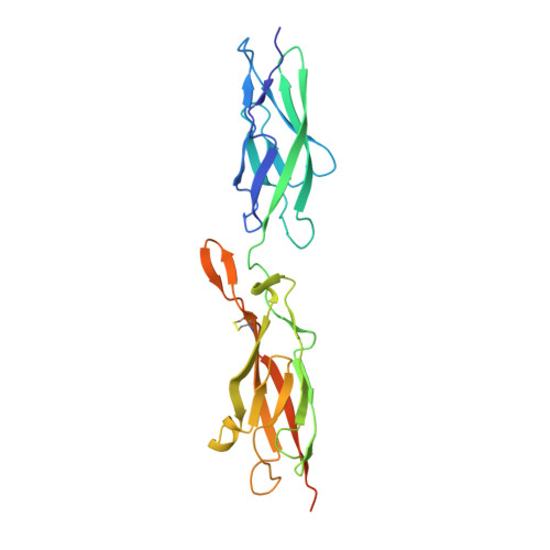
    Enterocytes are specialized epithelial cells lining the luminal surface of the small intestine that build densely packed arrays of microvilli known as brush borders. These microvilli drive nutrient absorption and are arranged in a hexagonal pattern maintained by intermicrovillar links formed by 2 nonclassical members of the cadherin superfamily of calcium-dependent cell adhesion proteins: protocadherin-24 (PCDH24, also known as CDHR2) and the mucin-like protocadherin (CDHR5). The extracellular domains of these proteins are involved in heterophilic and homophilic interactions important for intermicrovillar function, yet the structural determinants of these interactions remain unresolved. Here, we present X-ray crystal structures of the PCDH24 and CDHR5 extracellular tips and analyze their species-specific features relevant for adhesive interactions. In parallel, we use binding assays to identify the PCDH24 and CDHR5 domains involved in both heterophilic and homophilic adhesion for human and mouse proteins. Our results suggest that homophilic and heterophilic interactions involving PCDH24 and CDHR5 are species dependent with unique and distinct minimal adhesive units.


  • Organizational Affiliation

    Ohio State Biochemistry Program, The Ohio State University, Columbus, Ohio, United States of America.


Macromolecules
Find similar proteins by:  (by identity cutoff)  |  3D Structure
Entity ID: 1
MoleculeChains Sequence LengthOrganismDetailsImage
Cadherin-related family member 2
A, B, C, D
238Homo sapiensMutation(s): 0 
Gene Names: CDHR2PCDH24PCLKC
UniProt & NIH Common Fund Data Resources
Find proteins for Q9BYE9 (Homo sapiens)
Explore Q9BYE9 
Go to UniProtKB:  Q9BYE9
PHAROS:  Q9BYE9
GTEx:  ENSG00000074276 
Entity Groups  
Sequence Clusters30% Identity50% Identity70% Identity90% Identity95% Identity100% Identity
UniProt GroupQ9BYE9
Sequence Annotations
Expand
  • Reference Sequence
Oligosaccharides

Help

Entity ID: 2
MoleculeChains Length2D Diagram Glycosylation3D Interactions
beta-D-mannopyranose-(1-4)-2-acetamido-2-deoxy-beta-D-glucopyranose-(1-4)-2-acetamido-2-deoxy-beta-D-glucopyranose
E, F, G, H
3N-Glycosylation
Glycosylation Resources
GlyTouCan:  G15407YE
GlyCosmos:  G15407YE
GlyGen:  G15407YE
Small Molecules
Ligands 5 Unique
IDChains Name / Formula / InChI Key2D Diagram3D Interactions
IOD
Query on IOD

Download Ideal Coordinates CCD File 
R [auth B]IODIDE ION
I
XMBWDFGMSWQBCA-UHFFFAOYSA-M
TRS
Query on TRS

Download Ideal Coordinates CCD File 
I [auth A]2-AMINO-2-HYDROXYMETHYL-PROPANE-1,3-DIOL
C4 H12 N O3
LENZDBCJOHFCAS-UHFFFAOYSA-O
CA
Query on CA

Download Ideal Coordinates CCD File 
BA [auth D]
CA [auth D]
DA [auth D]
EA [auth D]
J [auth A]
BA [auth D],
CA [auth D],
DA [auth D],
EA [auth D],
J [auth A],
K [auth A],
L [auth A],
O [auth B],
P [auth B],
Q [auth B],
U [auth C],
V [auth C],
W [auth C],
X [auth C]
CALCIUM ION
Ca
BHPQYMZQTOCNFJ-UHFFFAOYSA-N
CL
Query on CL

Download Ideal Coordinates CCD File 
AA [auth C]
FA [auth D]
M [auth A]
S [auth B]
Y [auth C]
AA [auth C],
FA [auth D],
M [auth A],
S [auth B],
Y [auth C],
Z [auth C]
CHLORIDE ION
Cl
VEXZGXHMUGYJMC-UHFFFAOYSA-M
NA
Query on NA

Download Ideal Coordinates CCD File 
N [auth A],
T [auth B]
SODIUM ION
Na
FKNQFGJONOIPTF-UHFFFAOYSA-N
Experimental Data & Validation

Experimental Data

  • Method: X-RAY DIFFRACTION
  • Resolution: 3.17 Å
  • R-Value Free: 0.290 
  • R-Value Work: 0.213 
  • Space Group: P 1 21 1
Unit Cell:
Length ( Å )Angle ( ˚ )
a = 63.899α = 90
b = 86.641β = 103.458
c = 104.892γ = 90
Software Package:
Software NamePurpose
REFMACrefinement
HKL-2000data reduction
HKL-2000data scaling
PHASERphasing

Structure Validation

View Full Validation Report


Entry History & Funding Information

Deposition Data


Funding OrganizationLocationGrant Number
National Institutes of Health/National Institute of Diabetes and Digestive and Kidney Disease (NIH/NIDDK)United StatesR01 DK095811

Revision History  (Full details and data files)

  • Version 1.0: 2021-09-01
    Type: Initial release
  • Version 1.1: 2021-12-22
    Changes: Database references
  • Version 1.2: 2023-10-18
    Changes: Data collection, Refinement description