7MWG

16-nm repeat microtubule doublet


Experimental Data Snapshot

  • Method: ELECTRON MICROSCOPY
  • Resolution: 3.50 Å
  • Aggregation State: FILAMENT 
  • Reconstruction Method: SINGLE PARTICLE 

wwPDB Validation   3D Report Full Report


This is version 1.2 of the entry. See complete history


Literature

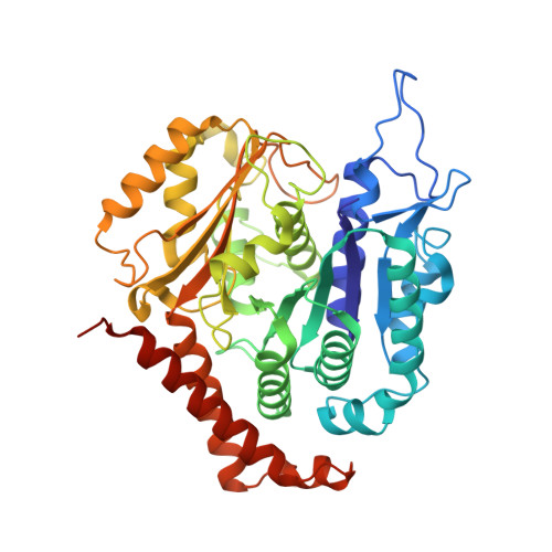
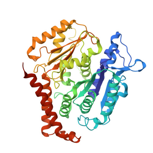
Structures of outer-arm dynein array on microtubule doublet reveal a motor coordination mechanism.

Rao, Q.Han, L.Wang, Y.Chai, P.Kuo, Y.W.Yang, R.Hu, F.Yang, Y.Howard, J.Zhang, K.

(2021) Nat Struct Mol Biol 28: 799-810

  • DOI: https://doi.org/10.1038/s41594-021-00656-9
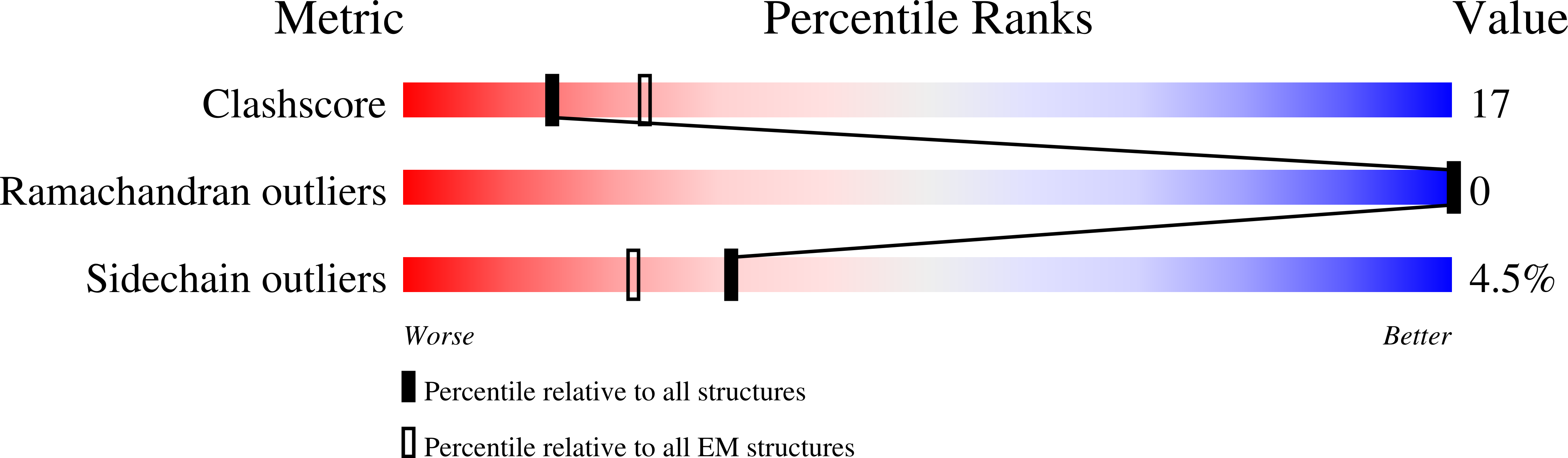
  • Primary Citation of Related Structures:  
    7K58, 7K5B, 7KEK, 7MWG, 7N32

  • PubMed Abstract: 

    Thousands of outer-arm dyneins (OADs) are arrayed in the axoneme to drive a rhythmic ciliary beat. Coordination among multiple OADs is essential for generating mechanical forces to bend microtubule doublets (MTDs). Using electron microscopy, we determined high-resolution structures of Tetrahymena thermophila OAD arrays bound to MTDs in two different states. OAD preferentially binds to MTD protofilaments with a pattern resembling the native tracks for its distinct microtubule-binding domains. Upon MTD binding, free OADs are induced to adopt a stable parallel conformation, primed for array formation. Extensive tail-to-head (TTH) interactions between OADs are observed, which need to be broken for ATP turnover by the dynein motor. We propose that OADs in an array sequentially hydrolyze ATP to slide the MTDs. ATP hydrolysis in turn relaxes the TTH interfaces to effect free nucleotide cycles of downstream OADs. These findings lead to a model explaining how conformational changes in the axoneme produce coordinated action of dyneins.


  • Organizational Affiliation

    Department of Molecular Biophysics and Biochemistry, Yale University, New Haven, CT, USA.


Macromolecules
Find similar proteins by:  (by identity cutoff)  |  3D Structure
Entity ID: 1
MoleculeChains Sequence LengthOrganismDetailsImage
Tubulin alpha chain449Tetrahymena thermophilaMutation(s): 0 
UniProt
Find proteins for P41351 (Tetrahymena thermophila)
Explore P41351 
Go to UniProtKB:  P41351
Entity Groups  
Sequence Clusters30% Identity50% Identity70% Identity90% Identity95% Identity100% Identity
UniProt GroupP41351
Sequence Annotations
Expand
  • Reference Sequence
Find similar proteins by:  (by identity cutoff)  |  3D Structure
Entity ID: 2
MoleculeChains Sequence LengthOrganismDetailsImage
Tubulin beta chain443Tetrahymena thermophilaMutation(s): 0 
UniProt
Find proteins for P41352 (Tetrahymena thermophila)
Explore P41352 
Go to UniProtKB:  P41352
Entity Groups  
Sequence Clusters30% Identity50% Identity70% Identity90% Identity95% Identity100% Identity
UniProt GroupP41352
Sequence Annotations
Expand
  • Reference Sequence
Small Molecules
Experimental Data & Validation

Experimental Data

  • Method: ELECTRON MICROSCOPY
  • Resolution: 3.50 Å
  • Aggregation State: FILAMENT 
  • Reconstruction Method: SINGLE PARTICLE 

Structure Validation

View Full Validation Report


Entry History 

Deposition Data

Revision History  (Full details and data files)

  • Version 1.0: 2021-09-29
    Type: Initial release
  • Version 1.1: 2021-10-06
    Changes: Database references
  • Version 1.2: 2021-10-20
    Changes: Database references