7MK8

Crystal structure of a surface mutant of human fetal-specific CYP3A7 bound to dithiothreitol


Experimental Data Snapshot

  • Method: X-RAY DIFFRACTION
  • Resolution: 2.15 Å
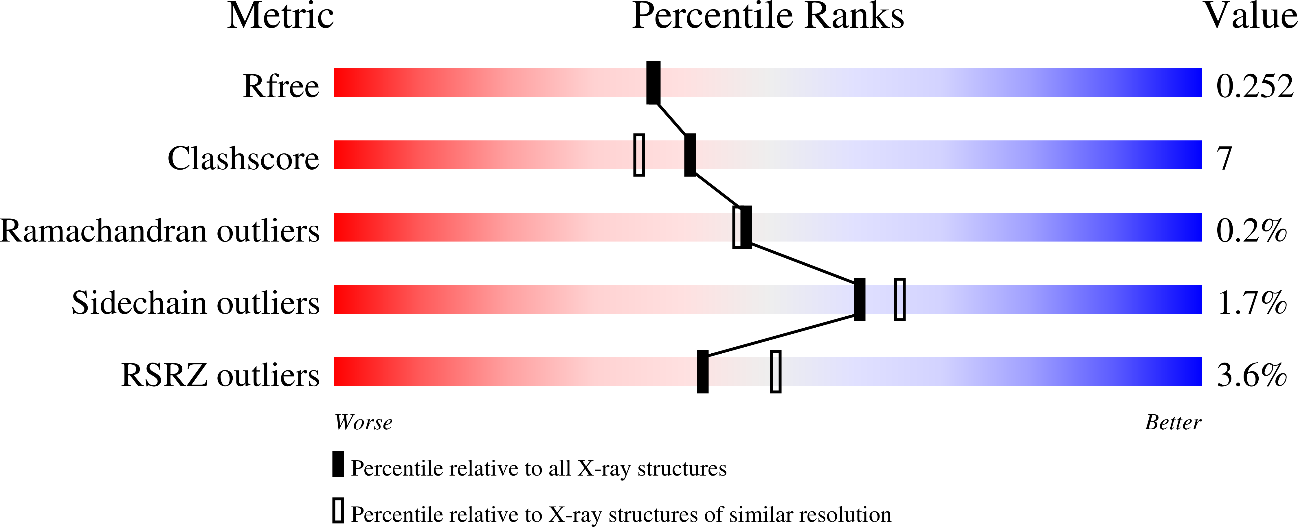
  • R-Value Free: 0.253 
  • R-Value Work: 0.203 
  • R-Value Observed: 0.205 

wwPDB Validation   3D Report Full Report


Ligand Structure Quality Assessment 


This is version 1.2 of the entry. See complete history


Literature

Structural Basis for the Diminished Ligand Binding and Catalytic Ability of Human Fetal-Specific CYP3A7.

Sevrioukova, I.F.

(2021) Int J Mol Sci 22

  • DOI: https://doi.org/10.3390/ijms22115831
  • Primary Citation of Related Structures:  
    7MK8

  • PubMed Abstract: 

    Cytochrome P450 3A7 (CYP3A7) is a fetal/neonatal liver enzyme that participates in estriol synthesis, clearance of all-trans retinoic acid, and xenobiotic metabolism. Compared to the closely related major drug-metabolizing enzyme in adult liver, CYP3A4, the ligand binding and catalytic capacity of CYP3A7 are substantially reduced. To better understand the structural basis for these functional differences, the 2.15 Å crystal structure of CYP3A7 has been solved. Comparative analysis of CYP3A enzymes shows that decreased structural plasticity rather than the active site microenvironment defines the ligand binding ability of CYP3A7. In particular, a rotameric switch in the gatekeeping amino acid F304 triggers local and long-range rearrangements that transmit to the F-G fragment and alter its interactions with the I-E-D-helical core, resulting in a more rigid structure. Elongation of the β 3 4 strands, H-bond linkage in the substrate channel, and steric constraints in the C-terminal loop further increase the active site rigidity and limit conformational ensemble. Collectively, these structural distinctions lower protein plasticity and change the heme environment, which, in turn, could impede the spin-state transition essential for optimal reactivity and oxidation of substrates.


  • Organizational Affiliation

    Department of Molecular Biology and Biochemistry, University of California, Irvine, CA 92697-3900, USA.


Macromolecules
Find similar proteins by:  (by identity cutoff)  |  3D Structure
Entity ID: 1
MoleculeChains Sequence LengthOrganismDetailsImage
Isoform 2 of Cytochrome P450 3A7
A, B
519Homo sapiensMutation(s): 6 
Gene Names: CYP3A7
EC: 1.14.14.1
Membrane Entity: Yes 
UniProt & NIH Common Fund Data Resources
Find proteins for P24462 (Homo sapiens)
Explore P24462 
Go to UniProtKB:  P24462
PHAROS:  P24462
GTEx:  ENSG00000160870 
Entity Groups  
Sequence Clusters30% Identity50% Identity70% Identity90% Identity95% Identity100% Identity
UniProt GroupP24462
Sequence Annotations
Expand
  • Reference Sequence
Experimental Data & Validation

Experimental Data

  • Method: X-RAY DIFFRACTION
  • Resolution: 2.15 Å
  • R-Value Free: 0.253 
  • R-Value Work: 0.203 
  • R-Value Observed: 0.205 
  • Space Group: C 2 2 21
Unit Cell:
Length ( Å )Angle ( ˚ )
a = 72.29α = 90
b = 205.12β = 90
c = 156.55γ = 90
Software Package:
Software NamePurpose
PHENIXrefinement
PDB_EXTRACTdata extraction
MOSFLMdata reduction
SCALAdata scaling
PHASERphasing

Structure Validation

View Full Validation Report


Ligand Structure Quality Assessment 


Entry History & Funding Information

Deposition Data


Funding OrganizationLocationGrant Number
National Institutes of Health/National Institute of Environmental Health Sciences (NIH/NIEHS)United StatesES025767

Revision History  (Full details and data files)

  • Version 1.0: 2021-06-02
    Type: Initial release
  • Version 1.1: 2021-07-14
    Changes: Database references
  • Version 1.2: 2023-10-18
    Changes: Data collection, Database references, Refinement description