7MEZ

Structure of the phosphoinositide 3-kinase p110 gamma (PIK3CG) p101 (PIK3R5) complex


Experimental Data Snapshot

  • Method: ELECTRON MICROSCOPY
  • Resolution: 2.89 Å
  • Aggregation State: PARTICLE 
  • Reconstruction Method: SINGLE PARTICLE 

wwPDB Validation   3D Report Full Report


This is version 1.1 of the entry. See complete history


Literature

Structure of the phosphoinositide 3-kinase (PI3K) p110 gamma-p101 complex reveals molecular mechanism of GPCR activation.

Rathinaswamy, M.K.Dalwadi, U.Fleming, K.D.Adams, C.Stariha, J.T.B.Pardon, E.Baek, M.Vadas, O.DiMaio, F.Steyaert, J.Hansen, S.D.Yip, C.K.Burke, J.E.

(2021) Sci Adv 7

  • DOI: https://doi.org/10.1126/sciadv.abj4282
  • Primary Citation of Related Structures:  
    7MEZ

  • PubMed Abstract: 

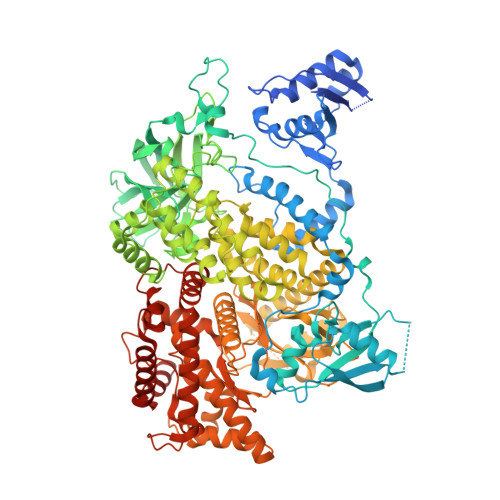
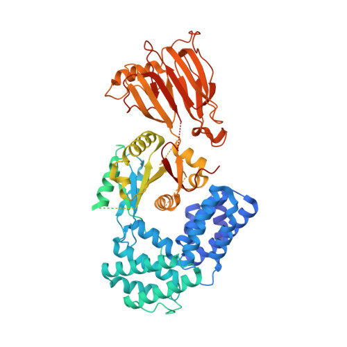
    The class IB phosphoinositide 3-kinase (PI3K), PI3Kγ, is a master regulator of immune cell function and a promising drug target for both cancer and inflammatory diseases. Critical to PI3Kγ function is the association of the p110γ catalytic subunit to either a p101 or p84 regulatory subunit, which mediates activation by G protein-coupled receptors. Here, we report the cryo-electron microscopy structure of a heterodimeric PI3Kγ complex, p110γ-p101. This structure reveals a unique assembly of catalytic and regulatory subunits that is distinct from other class I PI3K complexes. p101 mediates activation through its Gβγ-binding domain, recruiting the heterodimer to the membrane and allowing for engagement of a secondary Gβγ-binding site in p110γ. Mutations at the p110γ-p101 and p110γ-adaptor binding domain interfaces enhanced Gβγ activation. A nanobody that specifically binds to the p101-Gβγ interface blocks activation, providing a novel tool to study and target p110γ-p101-specific signaling events in vivo.


  • Organizational Affiliation

    Department of Biochemistry and Microbiology, University of Victoria, Victoria, British Columbia, Canada.


Macromolecules
Find similar proteins by:  (by identity cutoff)  |  3D Structure
Entity ID: 1
MoleculeChains Sequence LengthOrganismDetailsImage
Phosphatidylinositol 4,5-bisphosphate 3-kinase catalytic subunit gamma isoform1,102Homo sapiensMutation(s): 0 
Gene Names: PIK3CG
EC: 2.7.1.137 (PDB Primary Data), 2.7.1.153 (PDB Primary Data), 2.7.1.154 (PDB Primary Data), 2.7.11.1 (PDB Primary Data)
UniProt & NIH Common Fund Data Resources
Find proteins for P48736 (Homo sapiens)
Explore P48736 
Go to UniProtKB:  P48736
PHAROS:  P48736
GTEx:  ENSG00000105851 
Entity Groups  
Sequence Clusters30% Identity50% Identity70% Identity90% Identity95% Identity100% Identity
UniProt GroupP48736
Sequence Annotations
Expand
  • Reference Sequence
Find similar proteins by:  (by identity cutoff)  |  3D Structure
Entity ID: 2
MoleculeChains Sequence LengthOrganismDetailsImage
Phosphoinositide 3-kinase regulatory subunit 5881Sus scrofaMutation(s): 2 
Gene Names: PIK3R5
UniProt
Find proteins for O02696 (Sus scrofa)
Explore O02696 
Go to UniProtKB:  O02696
Entity Groups  
Sequence Clusters30% Identity50% Identity70% Identity90% Identity95% Identity100% Identity
UniProt GroupO02696
Sequence Annotations
Expand
  • Reference Sequence
Experimental Data & Validation

Experimental Data

  • Method: ELECTRON MICROSCOPY
  • Resolution: 2.89 Å
  • Aggregation State: PARTICLE 
  • Reconstruction Method: SINGLE PARTICLE 
EM Software:
TaskSoftware PackageVersion
RECONSTRUCTIONcryoSPARC3.0

Structure Validation

View Full Validation Report


Entry History & Funding Information

Deposition Data


Funding OrganizationLocationGrant Number
Canadian Institutes of Health Research (CIHR)Canada168998

Revision History  (Full details and data files)

  • Version 1.0: 2021-07-14
    Type: Initial release
  • Version 1.1: 2022-04-06
    Changes: Database references