Structural and biochemical studies of an iterative ribosomal peptide macrocyclase.
Li, G., Patel, K., Zhang, Y., Pugmire, J.K., Ding, Y., Bruner, S.D.(2022) Proteins 90: 670-679
- PubMed: 34664307
- DOI: https://doi.org/10.1002/prot.26264
- Primary Citation of Related Structures:
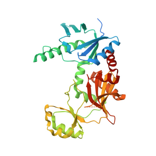
7M4S - PubMed Abstract:
Microviridins, tricyclic peptide natural products originally isolated from cyanobacteria, function as inhibitors of diverse serine-type proteases. Here we report the structure and biochemical characterization of AMdnB, a unique iterative macrocyclase involved in a microviridin biosynthetic pathway from Anabaena sp. PCC 7120. The ATP-dependent cyclase, along with the homologous AMdnC, introduce up to nine macrocyclizations on three distinct core regions of a precursor peptide, AMdnA. The results presented here provide structural and mechanistic insight into the iterative chemistry of AMdnB. In vitro AMdnB-catalyzed cyclization reactions demonstrate the synthesis of the two predicted tricyclic products from a multi-core precursor peptide substrate, consistent with a distributive mode of catalysis. The X-ray structure of AMdnB shows a structural motif common to ATP-grasp cyclases involved in RiPPs biosynthesis. Additionally, comparison with the noniterative MdnB allows insight into the structural basis for the iterative chemistry. Overall, the presented results provide insight into the general mechanism of iterative enzymes in ribosomally synthesized and post-translationally modified peptide biosynthetic pathways.
Organizational Affiliation:
Department of Chemistry, University of Florida, Gainesville, Florida, USA.