7M15

crystal structure of cj1430 in the presence of GDP-D-glycero-L-gluco-heptose, a GDP-D-glycero-4-keto-D-lyxo-heptose-3,5-epimerase from campylobacter jejuni


Experimental Data Snapshot

  • Method: X-RAY DIFFRACTION
  • Resolution: 1.85 Å
  • R-Value Free: 0.234 
  • R-Value Work: 0.190 
  • R-Value Observed: 0.192 

wwPDB Validation   3D Report Full Report


Ligand Structure Quality Assessment 


This is version 1.3 of the entry. See complete history


Literature

Biosynthesis of d- glycero -l- gluco -Heptose in the Capsular Polysaccharides of Campylobacter jejuni .

Huddleston, J.P.Anderson, T.K.Girardi, N.M.Thoden, J.B.Taylor, Z.Holden, H.M.Raushel, F.M.

(2021) Biochemistry 60: 1552-1563

  • DOI: https://doi.org/10.1021/acs.biochem.1c00183
  • Primary Citation of Related Structures:  
    7M13, 7M14, 7M15

  • PubMed Abstract: 

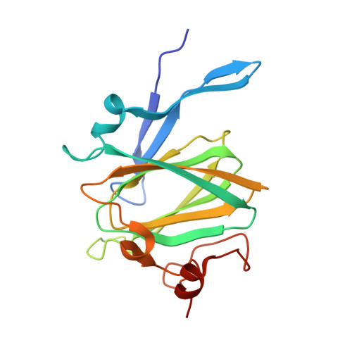
    Campylobacter jejuni is the leading cause of food poisoning in the United States and Europe. The exterior cell surface of C. jejuni is coated with a capsular polysaccharide (CPS) that is essential for the maintenance and integrity of the bacterial cell wall and evasion of the host immune response. The identity and sequences of the monosaccharide components of the CPS are quite variable and dependent on the specific strain of C. jejuni . It is currently thought that the immediate precursor for the multiple variations found in the heptose moieties of the C. jejuni CPS is GDP-d- glycero -α-d- manno -heptose. In C. jejuni NCTC 11168, the heptose moiety is d- glycero -l- gluco -heptose. It has previously been shown that Cj1427 catalyzes the oxidation of GDP-d- glycero -α-d- manno -heptose to GDP-d- glycero -4-keto-α-d- lyxo -heptose using α-ketoglutarate as a cosubstrate. Cj1430 was now demonstrated to catalyze the double epimerization of this product at C3 and C5 to form GDP-d- glycero -4-keto-β-l- xylo -heptose. Cj1428 subsequently catalyzes the stereospecific reduction of this GDP-linked heptose by NADPH to form GDP-d- glycero -β-l- gluco -heptose. The three-dimensional crystal structure of Cj1430 was determined to a resolution of 1.85 Å in the presence of bound GDP-d- glycero -β-l- gluco -heptose, a product analogue. The structure shows that it belongs to the cupin superfamily. The three-dimensional crystal structure of Cj1428 was solved in the presence of NADPH to a resolution of 1.50 Å. Its fold places it into the short-chain dehydrogenase/reductase superfamily. Typically, members in this family display a characteristic signature sequence of YXXXK, with the conserved tyrosine serving a key role in catalysis. In Cj1428, this residue is a phenylalanine.


  • Organizational Affiliation

    Department of Chemistry, Texas A&M University, College Station, Texas 77843, United States.


Macromolecules
Find similar proteins by:  (by identity cutoff)  |  3D Structure
Entity ID: 1
MoleculeChains Sequence LengthOrganismDetailsImage
GDP-D-glycero-L-gluco-heptose
A, B, C, D, E
A, B, C, D, E, F
185Campylobacter jejuni subsp. jejuni NCTC 11168 = ATCC 700819Mutation(s): 2 
Gene Names: rfbCCj1430c
EC: 5.1.3.13
UniProt
Find proteins for Q0P8I4 (Campylobacter jejuni subsp. jejuni serotype O:2 (strain ATCC 700819 / NCTC 11168))
Explore Q0P8I4 
Go to UniProtKB:  Q0P8I4
Entity Groups  
Sequence Clusters30% Identity50% Identity70% Identity90% Identity95% Identity100% Identity
UniProt GroupQ0P8I4
Sequence Annotations
Expand
  • Reference Sequence
Small Molecules
Ligands 2 Unique
IDChains Name / Formula / InChI Key2D Diagram3D Interactions
YO7
Query on YO7

Download Ideal Coordinates CCD File 
G [auth A]
H [auth B]
J [auth C]
L [auth D]
M [auth E]
G [auth A],
H [auth B],
J [auth C],
L [auth D],
M [auth E],
P [auth F]
[(2R,3S,4R,5R)-5-(2-amino-6-oxo-1,6-dihydro-9H-purin-9-yl)-3,4-dihydroxyoxolan-2-yl]methyl (2R,3S,4R,5R,6S)-6-[(1R)-1,2-dihydroxyethyl]-3,4,5-trihydroxyoxan-2-yl dihydrogen diphosphate (non-preferred name)
C17 H27 N5 O17 P2
HRUXIQZFNLQZQA-GVGRRHNDSA-N
EDO
Query on EDO

Download Ideal Coordinates CCD File 
I [auth B],
K [auth C],
N [auth E],
O [auth E],
Q [auth F]
1,2-ETHANEDIOL
C2 H6 O2
LYCAIKOWRPUZTN-UHFFFAOYSA-N
Experimental Data & Validation

Experimental Data

  • Method: X-RAY DIFFRACTION
  • Resolution: 1.85 Å
  • R-Value Free: 0.234 
  • R-Value Work: 0.190 
  • R-Value Observed: 0.192 
  • Space Group: C 1 2 1
Unit Cell:
Length ( Å )Angle ( ˚ )
a = 136.409α = 90
b = 81.569β = 94.38
c = 109.453γ = 90
Software Package:
Software NamePurpose
REFMACrefinement
PDB_EXTRACTdata extraction
SAINTdata reduction
SADABSdata scaling
PHASERphasing

Structure Validation

View Full Validation Report


Ligand Structure Quality Assessment 


Entry History & Funding Information

Deposition Data


Funding OrganizationLocationGrant Number
National Institutes of Health/National Institute of General Medical Sciences (NIH/NIGMS)United StatesGM122825
National Institutes of Health/National Institute of General Medical Sciences (NIH/NIGMS)United StatesGM134643
Robert A. Welch FoundationUnited StatesA-840

Revision History  (Full details and data files)

  • Version 1.0: 2021-03-24
    Type: Initial release
  • Version 1.1: 2021-05-12
    Changes: Database references
  • Version 1.2: 2021-06-02
    Changes: Database references
  • Version 1.3: 2023-10-18
    Changes: Data collection, Database references, Refinement description