7LXF

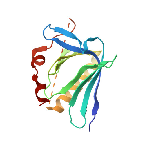
ENAH EVH1 domain bound to peptide from protein PCARE


Experimental Data Snapshot

  • Method: X-RAY DIFFRACTION
  • Resolution: 1.65 Å
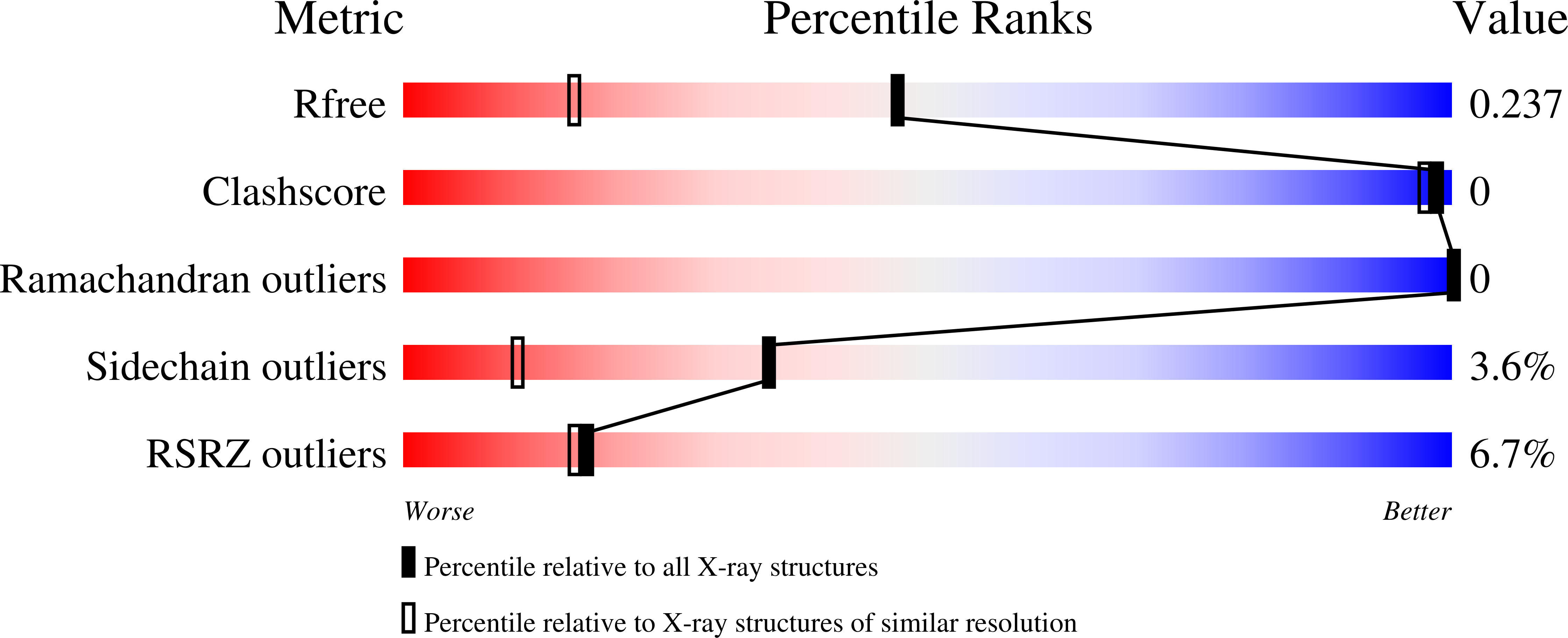
  • R-Value Free: 0.234 
  • R-Value Work: 0.217 
  • R-Value Observed: 0.218 

wwPDB Validation   3D Report Full Report


This is version 1.2 of the entry. See complete history


Literature

A distributed residue network permits conformational binding specificity in a conserved family of actin remodelers.

Hwang, T.Parker, S.S.Hill, S.M.Ilunga, M.W.Grant, R.A.Mouneimne, G.Keating, A.E.

(2021) Elife 10

  • DOI: https://doi.org/10.7554/eLife.70601
  • Primary Citation of Related Structures:  
    7LXF

  • PubMed Abstract: 

    Metazoan proteomes contain many paralogous proteins that have evolved distinct functions. The Ena/VASP family of actin regulators consists of three members that share an EVH1 interaction domain with a 100 % conserved binding site. A proteome-wide screen revealed photoreceptor cilium actin regulator (PCARE) as a high-affinity ligand for ENAH EVH1. Here, we report the surprising observation that PCARE is ~100-fold specific for ENAH over paralogs VASP and EVL and can selectively bind ENAH and inhibit ENAH-dependent adhesion in cells. Specificity arises from a mechanism whereby PCARE stabilizes a conformation of the ENAH EVH1 domain that is inaccessible to family members VASP and EVL. Structure-based modeling rapidly identified seven residues distributed throughout EVL that are sufficient to differentiate binding by ENAH vs. EVL. By exploiting the ENAH-specific conformation, we rationally designed the tightest and most selective ENAH binder to date. Our work uncovers a conformational mechanism of interaction specificity that distinguishes highly similar paralogs and establishes tools for dissecting specific Ena/VASP functions in processes including cancer cell invasion.


  • Organizational Affiliation

    Department of Biology, Massachusetts Institute of Technology, Cambridge, United States.


Macromolecules
Find similar proteins by:  (by identity cutoff)  |  3D Structure
Entity ID: 1
MoleculeChains Sequence LengthOrganismDetailsImage
Protein enabled homolog,Photoreceptor cilium actin regulator156Homo sapiensMutation(s): 0 
Gene Names: ENAHPCAREC2orf71
UniProt & NIH Common Fund Data Resources
Find proteins for A6NGG8 (Homo sapiens)
Explore A6NGG8 
Go to UniProtKB:  A6NGG8
PHAROS:  A6NGG8
GTEx:  ENSG00000179270 
Entity Groups  
Sequence Clusters30% Identity50% Identity70% Identity90% Identity95% Identity100% Identity
UniProt GroupA6NGG8
Sequence Annotations
Expand
  • Reference Sequence
Experimental Data & Validation

Experimental Data

  • Method: X-RAY DIFFRACTION
  • Resolution: 1.65 Å
  • R-Value Free: 0.234 
  • R-Value Work: 0.217 
  • R-Value Observed: 0.218 
  • Space Group: P 61 2 2
Unit Cell:
Length ( Å )Angle ( ˚ )
a = 52.12α = 90
b = 52.12β = 90
c = 197.85γ = 120
Software Package:
Software NamePurpose
PHENIXrefinement
XSCALEdata scaling
PDB_EXTRACTdata extraction
XDSdata reduction
PHASERphasing

Structure Validation

View Full Validation Report


Entry History & Funding Information

Deposition Data


Funding OrganizationLocationGrant Number
National Institutes of Health/National Institute of General Medical Sciences (NIH/NIGMS)United StatesGM129007-03

Revision History  (Full details and data files)

  • Version 1.0: 2021-11-24
    Type: Initial release
  • Version 1.1: 2022-06-08
    Changes: Database references
  • Version 1.2: 2023-10-18
    Changes: Data collection, Refinement description