


Crystal Structure of Tetur04g02350
Danehsian, L., Kluza, A., Dermauw, W., Wybouw, N., Van Leeuwen, T., Chruszcz, M.To be published.
Experimental Data Snapshot
Starting Model: experimental
View more details
Entity ID: 1 | |||||
|---|---|---|---|---|---|
| Molecule | Chains | Sequence Length | Organism | Details | Image |
| UDP-glycosyltransferase 203A2 | 461 | Tetranychus urticae | Mutation(s): 0 Gene Names: 107359436, UGT203A2 | 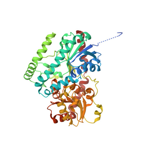 | |
UniProt | |||||
Find proteins for T1K1R5 (Tetranychus urticae) Explore T1K1R5 Go to UniProtKB: T1K1R5 | |||||
Entity Groups | |||||
| Sequence Clusters | 30% Identity50% Identity70% Identity90% Identity95% Identity100% Identity | ||||
| UniProt Group | T1K1R5 | ||||
Sequence AnnotationsExpand | |||||
| |||||
| Ligands 2 Unique | |||||
|---|---|---|---|---|---|
| ID | Chains | Name / Formula / InChI Key | 2D Diagram | 3D Interactions | |
| UDP (Subject of Investigation/LOI) Query on UDP | B [auth A] | URIDINE-5'-DIPHOSPHATE C9 H14 N2 O12 P2 XCCTYIAWTASOJW-XVFCMESISA-N |  | ||
| BGC (Subject of Investigation/LOI) Query on BGC | C [auth A], D [auth A] | beta-D-glucopyranose C6 H12 O6 WQZGKKKJIJFFOK-VFUOTHLCSA-N |  | ||
| Length ( Å ) | Angle ( ˚ ) |
|---|---|
| a = 111.159 | α = 90 |
| b = 61.596 | β = 106.15 |
| c = 78.861 | γ = 90 |
| Software Name | Purpose |
|---|---|
| REFMAC | refinement |
| PDB_EXTRACT | data extraction |
| HKL-3000 | data reduction |
| HKL-3000 | data scaling |
| MOLREP | phasing |
| Funding Organization | Location | Grant Number |
|---|---|---|
| United States Department of Agriculture (USDA) | United States | 2020-67014-31179 |
RCSB PDB (citation) is hosted by
RCSB PDB is a member of the

