7LRG

Filamentous actin decorated with human cardiac myosin binding protein C C2 domain

  • Classification: MOTOR PROTEIN
  • Organism(s): Sus scrofa, Homo sapiens
  • Expression System: Escherichia coli
  • Mutation(s): No 

  • Deposited: 2021-02-16 Released: 2021-08-11 
  • Deposition Author(s): Galkin, V.E.
  • Funding Organization(s): National Institutes of Health/National Heart, Lung, and Blood Institute (NIH/NHLBI), National Institutes of Health/National Institute of General Medical Sciences (NIH/NIGMS)

Experimental Data Snapshot

  • Method: ELECTRON MICROSCOPY
  • Resolution: 6.10 Å
  • Aggregation State: HELICAL ARRAY 
  • Reconstruction Method: HELICAL 

wwPDB Validation   3D Report Full Report


This is version 1.1 of the entry. See complete history


Literature

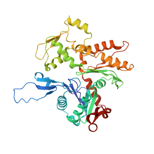
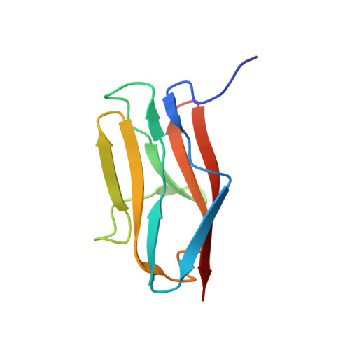
Interaction of the C2 Ig-like Domain of Cardiac Myosin Binding Protein-C with F-actin.

Risi, C.M.Patra, M.Belknap, B.Harris, S.P.White, H.D.Galkin, V.E.

(2021) J Mol Biol 433: 167178-167178

  • DOI: https://doi.org/10.1016/j.jmb.2021.167178
  • Primary Citation of Related Structures:  
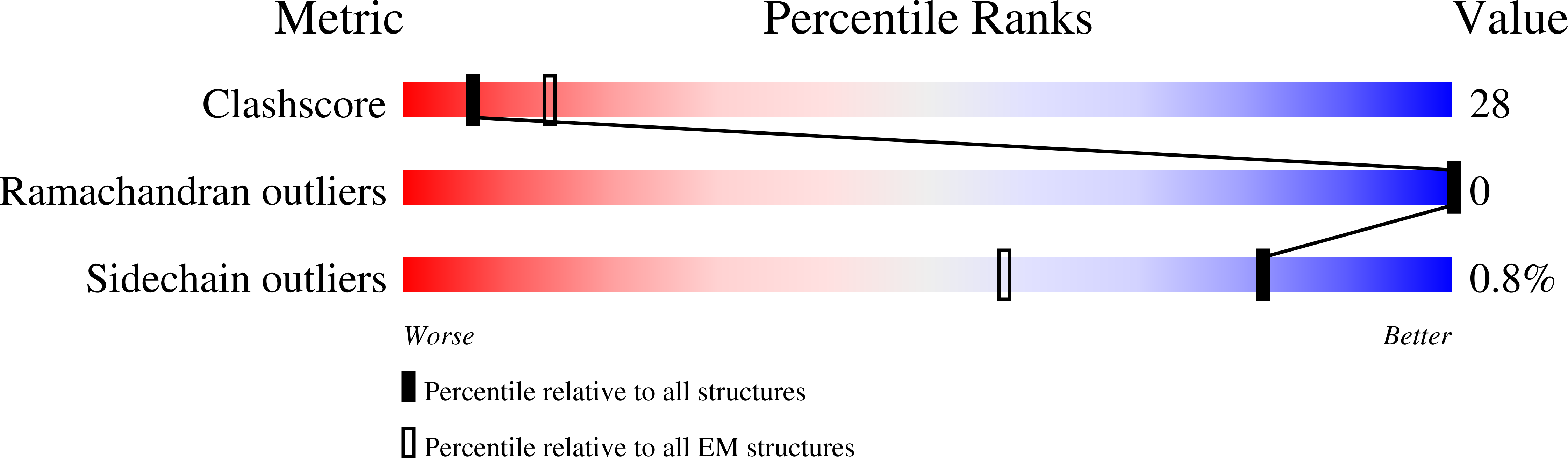
    7LRG

  • PubMed Abstract: 

    Cardiac muscle contraction depends on interactions between thick (myosin) and thin (actin) filaments (TFs). TFs are regulated by intracellular Ca 2+ levels. Under activating conditions Ca 2+ binds to the troponin complex and displaces tropomyosin from myosin binding sites on the TF surface to allow actomyosin interactions. Recent studies have shown that in addition to Ca 2+ , the first four N-terminal domains (NTDs) of cardiac myosin binding protein C (cMyBP-C) (e.g. C0, C1, M and C2), are potent modulators of the TF activity, but the mechanism of their collective action is poorly understood. Previously, we showed that C1 activates the TF at low Ca 2+ and C0 stabilizes binding of C1 to the TF, but the ability of C2 to bind and/or affect the TF remains unknown. Here we obtained 7.5 Å resolution cryo-EM reconstruction of C2-decorated actin filaments to demonstrate that C2 binds to actin in a single structural mode that does not activate the TF unlike the polymorphic binding of C0 and C1 to actin. Comparison of amino acid sequences of C2 with either C0 or C1 shows low levels of identity between the residues involved in interactions with the TF but high levels of conservation for residues involved in Ig fold stabilization. This provides a structural basis for strikingly different interactions of structurally homologous C0, C1 and C2 with the TF. Our detailed analysis of the interaction of C2 with the actin filament provides crucial information required to model the collective action of cMyBP-C NTDs on the cardiac TF.


  • Organizational Affiliation

    Department of Physiological Sciences, Eastern Virginia Medical School, Norfolk, VA 23507, USA.


Macromolecules
Find similar proteins by:  (by identity cutoff)  |  3D Structure
Entity ID: 1
MoleculeChains Sequence LengthOrganismDetailsImage
Actin, alpha cardiac muscle 1
A, B, C, D, E
A, B, C, D, E, F
377Sus scrofaMutation(s): 0 
UniProt
Find proteins for B6VNT8 (Sus scrofa)
Explore B6VNT8 
Go to UniProtKB:  B6VNT8
Entity Groups  
Sequence Clusters30% Identity50% Identity70% Identity90% Identity95% Identity100% Identity
UniProt GroupB6VNT8
Sequence Annotations
Expand
  • Reference Sequence
Find similar proteins by:  (by identity cutoff)  |  3D Structure
Entity ID: 2
MoleculeChains Sequence LengthOrganismDetailsImage
Myosin-binding protein C, cardiac-type
G, H, I, J, K
G, H, I, J, K, L
94Homo sapiensMutation(s): 0 
Gene Names: MYBPC3
UniProt & NIH Common Fund Data Resources
Find proteins for Q14896 (Homo sapiens)
Explore Q14896 
Go to UniProtKB:  Q14896
PHAROS:  Q14896
GTEx:  ENSG00000134571 
Entity Groups  
Sequence Clusters30% Identity50% Identity70% Identity90% Identity95% Identity100% Identity
UniProt GroupQ14896
Sequence Annotations
Expand
  • Reference Sequence
Experimental Data & Validation

Experimental Data

  • Method: ELECTRON MICROSCOPY
  • Resolution: 6.10 Å
  • Aggregation State: HELICAL ARRAY 
  • Reconstruction Method: HELICAL 
EM Software:
TaskSoftware PackageVersion
RECONSTRUCTIONIHRSR
MODEL REFINEMENTPHENIX

Structure Validation

View Full Validation Report


Entry History & Funding Information

Deposition Data

  • Released Date: 2021-08-11 
  • Deposition Author(s): Galkin, V.E.

Funding OrganizationLocationGrant Number
National Institutes of Health/National Heart, Lung, and Blood Institute (NIH/NHLBI)United StatesHL140925
National Institutes of Health/National Institute of General Medical Sciences (NIH/NIGMS)United StatesGM116790
National Institutes of Health/National Institute of General Medical Sciences (NIH/NIGMS)United StatesGM116788

Revision History  (Full details and data files)

  • Version 1.0: 2021-08-11
    Type: Initial release
  • Version 1.1: 2021-08-25
    Changes: Database references