7LQ6

CryoEM structure of Escherichia coli PBP1b


Experimental Data Snapshot

  • Method: ELECTRON MICROSCOPY
  • Resolution: 3.28 Å
  • Aggregation State: PARTICLE 
  • Reconstruction Method: SINGLE PARTICLE 

wwPDB Validation   3D Report Full Report


This is version 1.1 of the entry. See complete history


Literature

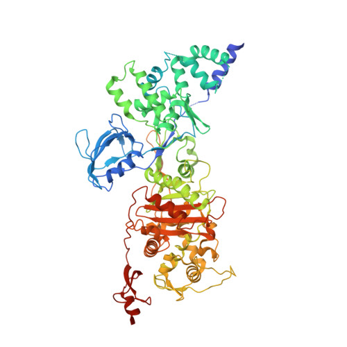
CryoEM structure of the antibacterial target PBP1b at 3.3 angstrom resolution.

Caveney, N.A.Workman, S.D.Yan, R.Atkinson, C.E.Yu, Z.Strynadka, N.C.J.

(2021) Nat Commun 12: 2775-2775

  • DOI: https://doi.org/10.1038/s41467-021-23063-6
  • Primary Citation of Related Structures:  
    7LQ6

  • PubMed Abstract: 

    The pathway for the biosynthesis of the bacterial cell wall is one of the most prolific antibiotic targets, exemplified by the widespread use of β-lactam antibiotics. Despite this, our structural understanding of class A penicillin binding proteins, which perform the last two steps in this pathway, is incomplete due to the inherent difficulty in their crystallization and the complexity of their substrates. Here, we determine the near atomic resolution structure of the 83 kDa class A PBP from Escherichia coli, PBP1b, using cryogenic electron microscopy and a styrene maleic acid anhydride membrane mimetic. PBP1b, in its apo form, is seen to exhibit a distinct conformation in comparison to Moenomycin-bound crystal structures. The work herein paves the way for the use of cryoEM in structure-guided antibiotic development for this notoriously difficult to crystalize class of proteins and their complex substrates.


  • Organizational Affiliation

    Department of Biochemistry and Molecular Biology, University of British Columbia, Vancouver, BC, Canada.


Macromolecules
Find similar proteins by:  (by identity cutoff)  |  3D Structure
Entity ID: 1
MoleculeChains Sequence LengthOrganismDetailsImage
Penicillin-binding protein 1B747Escherichia coli K-12Mutation(s): 0 
Gene Names: mrcBpbpFponBb0149JW0145
EC: 2.4.1.129 (PDB Primary Data), 3.4.16.4 (PDB Primary Data)
Membrane Entity: Yes 
UniProt
Find proteins for P02919 (Escherichia coli (strain K12))
Explore P02919 
Go to UniProtKB:  P02919
Entity Groups  
Sequence Clusters30% Identity50% Identity70% Identity90% Identity95% Identity100% Identity
UniProt GroupP02919
Sequence Annotations
Expand
  • Reference Sequence
Experimental Data & Validation

Experimental Data

  • Method: ELECTRON MICROSCOPY
  • Resolution: 3.28 Å
  • Aggregation State: PARTICLE 
  • Reconstruction Method: SINGLE PARTICLE 
EM Software:
TaskSoftware PackageVersion
RECONSTRUCTIONRELION
RECONSTRUCTIONcryoSPARC

Structure Validation

View Full Validation Report


Entry History & Funding Information

Deposition Data


Funding OrganizationLocationGrant Number
Canadian Institutes of Health Research (CIHR)Canada--

Revision History  (Full details and data files)

  • Version 1.0: 2021-05-26
    Type: Initial release
  • Version 1.1: 2024-03-06
    Changes: Data collection, Database references