7LFJ

MODEL OF MHC CLASS Ib H2-M3 WITH MOUSE ND1 N-TERMINAL HEPTAPEPTIDE, ALA MUTANT, REFINED AT 1.70 ANGSTROMS RESOLUTION


Experimental Data Snapshot

  • Method: X-RAY DIFFRACTION
  • Resolution: 1.70 Å
  • R-Value Free: 0.207 
  • R-Value Work: 0.175 
  • R-Value Observed: 0.176 

wwPDB Validation   3D Report Full Report


Ligand Structure Quality Assessment 


This is version 1.2 of the entry. See complete history


Literature

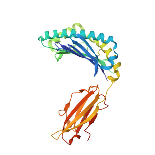
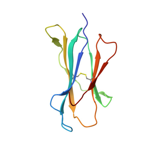
Structure and dynamics of major histocompatibility class Ib molecule H2-M3 complexed with mitochondrial-derived peptides.

Strand, A.Shen, S.T.Tomchick, D.R.Wang, J.Wang, C.R.Deisenhofer, J.

(2022) J Biomol Struct Dyn 40: 10300-10312

  • DOI: https://doi.org/10.1080/07391102.2021.1942214
  • Primary Citation of Related Structures:  
    7LFI, 7LFJ, 7LFK, 7LFL, 7LFM

  • PubMed Abstract: 

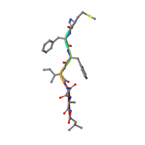
    Presentation of antigenic peptides to T-cell receptors is an essential step in the adaptive immune response. In the mouse the class Ib major histocompatibility complex molecule, H2-M3, presents bacterial- and mitochondrial-derived peptides to T-cell receptors on cytotoxic T cells. Four mitochondrial heptapeptides, differing only at residue 6, form complexes with H2-M3 which can be distinguished by T cells. No structures of relevant receptors are available. To investigate the structural basis for this distinction, crystal structures were determined and molecular dynamics simulations over one microsecond were done for each complex. In the crystal structures of the heptapeptide complexes with H2-M3, presented here, the side chains of the peptide residues at position 6 all point into the H2-M3 binding groove, and are thus inaccessible, so that the very similar structures do not suggest how recognition and initiation of responses by the T cells may occur. However, conformational differences, which could be crucial to T-cell discrimination, appear within one microsecond during molecular dynamics simulations of the four complexes. Specifically, the three C-terminal residues of peptide ligands with alanine or threonine at position 6 partially exit the binding groove; this does not occur in peptide ligands with isoleucine or valine at position 6. Structural changes associated with partial peptide exit from the binding groove, along with relevant peptide binding energetics and immunological results are discussed. Communicated by Ramaswamy H. Sarma.


  • Organizational Affiliation

    Green Center for Systems Biology, UT Southwestern Medical Center, Dallas, TX, USA.


Macromolecules
Find similar proteins by:  (by identity cutoff)  |  3D Structure
Entity ID: 1
MoleculeChains Sequence LengthOrganismDetailsImage
Histocompatibility 2, M region locus 3
A, D
282Mus musculusMutation(s): 0 
Gene Names: H2-M3H-2M3
UniProt
Find proteins for Q31093 (Mus musculus)
Explore Q31093 
Go to UniProtKB:  Q31093
Entity Groups  
Sequence Clusters30% Identity50% Identity70% Identity90% Identity95% Identity100% Identity
UniProt GroupQ31093
Sequence Annotations
Expand
  • Reference Sequence
Find similar proteins by:  (by identity cutoff)  |  3D Structure
Entity ID: 2
MoleculeChains Sequence LengthOrganismDetailsImage
Beta-2-microglobulin
B, E
99Mus musculusMutation(s): 1 
Gene Names: B2m
UniProt & NIH Common Fund Data Resources
Find proteins for P01887 (Mus musculus)
Explore P01887 
Go to UniProtKB:  P01887
IMPC:  MGI:88127
Entity Groups  
Sequence Clusters30% Identity50% Identity70% Identity90% Identity95% Identity100% Identity
UniProt GroupP01887
Sequence Annotations
Expand
  • Reference Sequence

Find similar proteins by:  Sequence   |   3D Structure  

Entity ID: 3
MoleculeChains Sequence LengthOrganismDetailsImage
Heptapeptide from NADH-ubiquinone oxidoreductase chain 1
C, F
7Mus musculusMutation(s): 1 
EC: 7.1.1.2
UniProt
Find proteins for P03888 (Mus musculus)
Explore P03888 
Go to UniProtKB:  P03888
Entity Groups  
UniProt GroupP03888
Sequence Annotations
Expand
  • Reference Sequence
Small Molecules
Modified Residues  1 Unique
IDChains TypeFormula2D DiagramParent
FME
Query on FME
C, F
L-PEPTIDE LINKINGC6 H11 N O3 SMET
Experimental Data & Validation

Experimental Data

  • Method: X-RAY DIFFRACTION
  • Resolution: 1.70 Å
  • R-Value Free: 0.207 
  • R-Value Work: 0.175 
  • R-Value Observed: 0.176 
  • Space Group: P 1
Unit Cell:
Length ( Å )Angle ( ˚ )
a = 63.866α = 99.435
b = 65.292β = 97.897
c = 55.125γ = 111.902
Software Package:
Software NamePurpose
PHENIXrefinement
PDB_EXTRACTdata extraction
HKL-2000data reduction
HKL-2000data scaling
PHENIXphasing

Structure Validation

View Full Validation Report


Ligand Structure Quality Assessment 


Entry History & Funding Information

Deposition Data


Funding OrganizationLocationGrant Number
National Institutes of Health/National Institute of General Medical Sciences (NIH/NIGMS)United StatesRO1-GM079383
National Institutes of Health/National Institute of General Medical Sciences (NIH/NIGMS)United StatesP30-DA035778

Revision History  (Full details and data files)

  • Version 1.0: 2021-07-14
    Type: Initial release
  • Version 1.1: 2022-12-28
    Changes: Database references
  • Version 1.2: 2023-10-25
    Changes: Data collection, Refinement description