7L4V

C-terminal bZIP domain of human C/EBPbeta Bound to DNA with Consensus Recognition with GT Mismatch


Experimental Data Snapshot

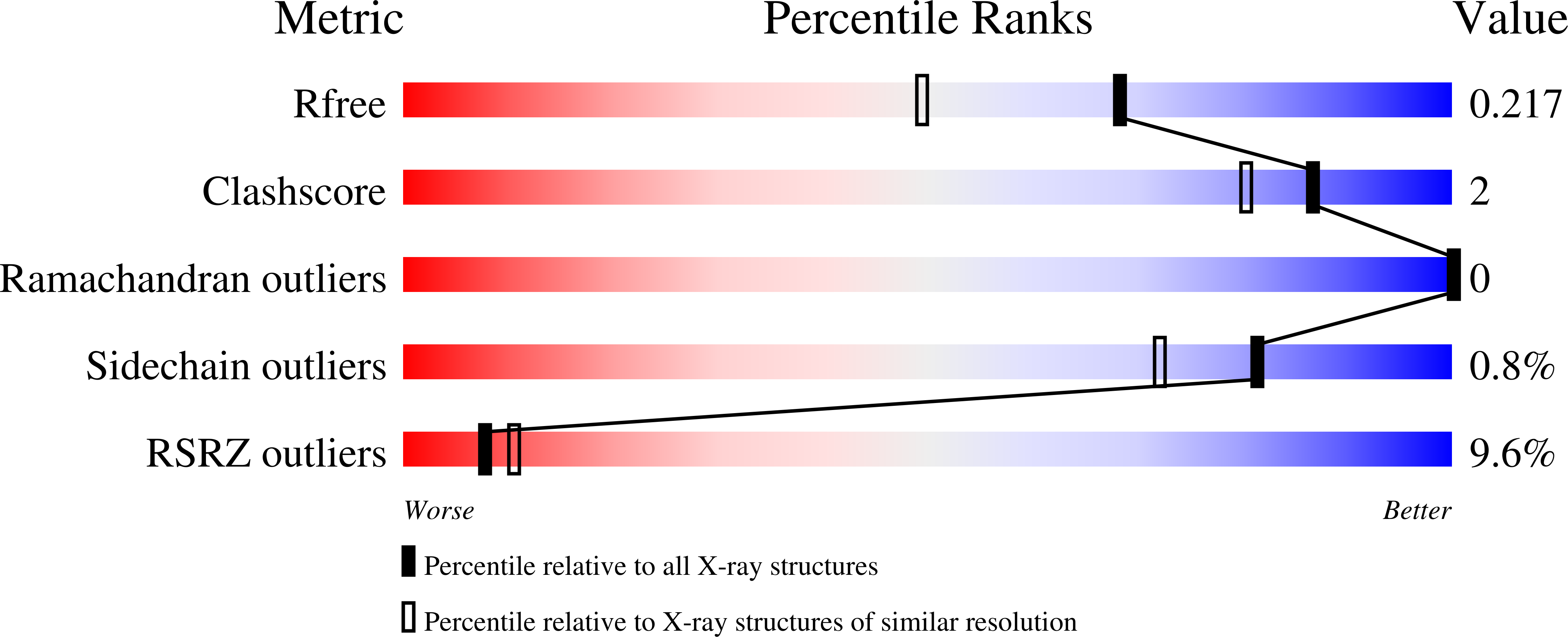
  • Method: X-RAY DIFFRACTION
  • Resolution: 1.75 Å
  • R-Value Free: 0.221 
  • R-Value Work: 0.199 
  • R-Value Observed: 0.200 

wwPDB Validation   3D Report Full Report


This is version 1.1 of the entry. See complete history


Literature

Preferential CEBP binding to T:G mismatches and increased C-to-T human somatic mutations.

Yang, J.Horton, J.R.Akdemir, K.C.Li, J.Huang, Y.Kumar, J.Blumenthal, R.M.Zhang, X.Cheng, X.

(2021) Nucleic Acids Res 49: 5084-5094

  • DOI: https://doi.org/10.1093/nar/gkab276
  • Primary Citation of Related Structures:  
    7L4V

  • PubMed Abstract: 

    DNA cytosine methylation in mammals modulates gene expression and chromatin accessibility. It also impacts mutation rates, via spontaneous oxidative deamination of 5-methylcytosine (5mC) to thymine. In most cases the resulting T:G mismatches are repaired, following T excision by one of the thymine DNA glycosylases, TDG or MBD4. We found that C-to-T mutations are enriched in the binding sites of CCAAT/enhancer binding proteins (CEBP). Within a CEBP site, the presence of a T:G mismatch increased CEBPβ binding affinity by a factor of >60 relative to the normal C:G base pair. This enhanced binding to a mismatch inhibits its repair by both TDG and MBD4 in vitro. Furthermore, repair of the deamination product of unmethylated cytosine, which yields a U:G DNA mismatch that is normally repaired via uracil DNA glycosylase, is also inhibited by CEBPβ binding. Passage of a replication fork over either a T:G or U:G mismatch, before repair can occur, results in a C-to-T mutation in one of the daughter duplexes. Our study thus provides a plausible mechanism for accumulation of C-to-T human somatic mutations.


  • Organizational Affiliation

    Department of Epigenetics and Molecular Carcinogenesis, University of Texas MD Anderson Cancer Center, Houston, TX 77030, USA.


Macromolecules

Find similar proteins by:  (by identity cutoff)  |  3D Structure
Entity ID: 1
MoleculeChains Sequence LengthOrganismDetailsImage
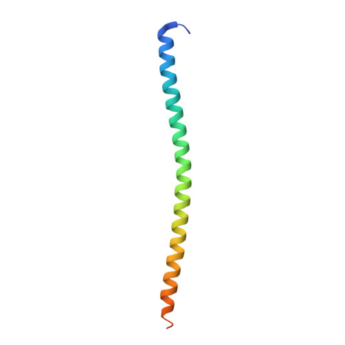
CCAAT/enhancer-binding protein beta
A, B
78Homo sapiensMutation(s): 0 
Gene Names: CEBPBTCF5PP9092
UniProt & NIH Common Fund Data Resources
Find proteins for P17676 (Homo sapiens)
Explore P17676 
Go to UniProtKB:  P17676
PHAROS:  P17676
GTEx:  ENSG00000172216 
Entity Groups  
Sequence Clusters30% Identity50% Identity70% Identity90% Identity95% Identity100% Identity
UniProt GroupP17676
Sequence Annotations
Expand
  • Reference Sequence

Find similar nucleic acids by:  Sequence   |   3D Structure  

Entity ID: 2
MoleculeChains LengthOrganismImage
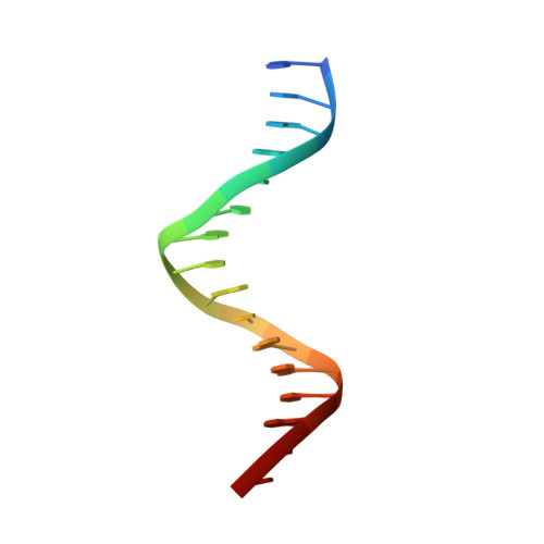
DNA Strand 116Homo sapiens
Sequence Annotations
Expand
  • Reference Sequence

Find similar nucleic acids by:  Sequence   |   3D Structure  

Entity ID: 3
MoleculeChains LengthOrganismImage
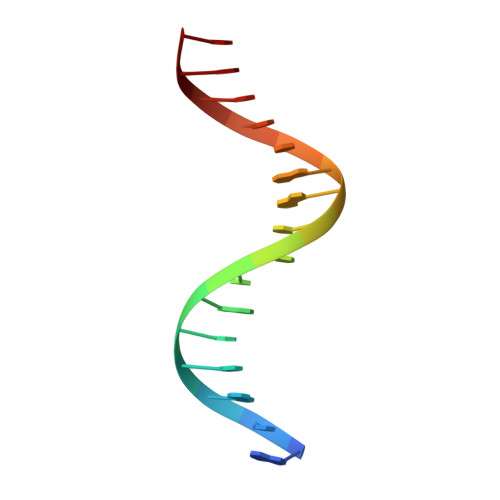
DNA Strand 216Homo sapiens
Sequence Annotations
Expand
  • Reference Sequence
Experimental Data & Validation

Experimental Data

  • Method: X-RAY DIFFRACTION
  • Resolution: 1.75 Å
  • R-Value Free: 0.221 
  • R-Value Work: 0.199 
  • R-Value Observed: 0.200 
  • Space Group: C 2 2 21
Unit Cell:
Length ( Å )Angle ( ˚ )
a = 101.452α = 90
b = 113.144β = 90
c = 75.74γ = 90
Software Package:
Software NamePurpose
PHENIXrefinement
SERGUIdata collection
HKL-2000data reduction
HKL-2000data scaling
PHENIXphasing

Structure Validation

View Full Validation Report


Entry History & Funding Information

Deposition Data


Funding OrganizationLocationGrant Number
National Institutes of Health/National Institute of General Medical Sciences (NIH/NIGMS)United StatesGM049245-23

Revision History  (Full details and data files)

  • Version 1.0: 2021-11-03
    Type: Initial release
  • Version 1.1: 2023-10-18
    Changes: Data collection, Refinement description