7L4I

Crystal structure of a substrate-trapping variant of PPM1H phosphatase


Experimental Data Snapshot

  • Method: X-RAY DIFFRACTION
  • Resolution: 2.58 Å
  • R-Value Free: 0.236 
  • R-Value Work: 0.190 
  • R-Value Observed: 0.192 

wwPDB Validation   3D Report Full Report


This is version 1.2 of the entry. See complete history


Literature

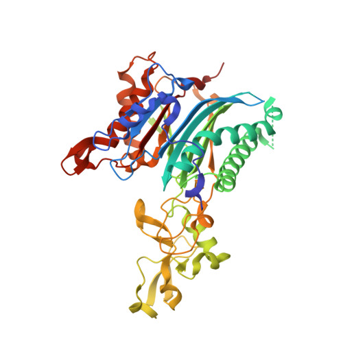
Structural basis for the specificity of PPM1H phosphatase for Rab GTPases.

Waschbusch, D.Berndsen, K.Lis, P.Knebel, A.Lam, Y.P.Alessi, D.R.Khan, A.R.

(2021) EMBO Rep 22: e52675-e52675

  • DOI: https://doi.org/10.15252/embr.202152675
  • Primary Citation of Related Structures:  
    7KPR, 7L4I, 7L4J, 7N0Z

  • PubMed Abstract: 

    LRRK2 serine/threonine kinase is associated with inherited Parkinson's disease. LRRK2 phosphorylates a subset of Rab GTPases within their switch 2 motif to control their interactions with effectors. Recent work has shown that the metal-dependent protein phosphatase PPM1H counteracts LRRK2 by dephosphorylating Rabs. PPM1H is highly selective for LRRK2 phosphorylated Rabs, and closely related PPM1J exhibits no activity towards substrates such as Rab8a phosphorylated at Thr72 (pThr72). Here, we have identified the molecular determinant of PPM1H specificity for Rabs. The crystal structure of PPM1H reveals a structurally conserved phosphatase fold that strikingly has evolved a 110-residue flap domain adjacent to the active site. The flap domain distantly resembles tudor domains that interact with histones in the context of epigenetics. Cellular assays, crosslinking and 3-D modelling suggest that the flap domain encodes the docking motif for phosphorylated Rabs. Consistent with this hypothesis, a PPM1J chimaera with the PPM1H flap domain dephosphorylates pThr72 of Rab8a both in vitro and in cellular assays. Therefore, PPM1H has acquired a Rab-specific interaction domain within a conserved phosphatase fold.


  • Organizational Affiliation

    School of Biochemistry and Immunology, Trinity College Dublin, Dublin 2, Ireland.


Macromolecules
Find similar proteins by:  (by identity cutoff)  |  3D Structure
Entity ID: 1
MoleculeChains Sequence LengthOrganismDetailsImage
Protein phosphatase 1H
A, B
451Homo sapiensMutation(s): 2 
Gene Names: PPM1HARHCL1KIAA1157URCC2
EC: 3.1.3.16
UniProt & NIH Common Fund Data Resources
Find proteins for Q9ULR3 (Homo sapiens)
Explore Q9ULR3 
Go to UniProtKB:  Q9ULR3
PHAROS:  Q9ULR3
GTEx:  ENSG00000111110 
Entity Groups  
Sequence Clusters30% Identity50% Identity70% Identity90% Identity95% Identity100% Identity
UniProt GroupQ9ULR3
Sequence Annotations
Expand
  • Reference Sequence
Experimental Data & Validation

Experimental Data

  • Method: X-RAY DIFFRACTION
  • Resolution: 2.58 Å
  • R-Value Free: 0.236 
  • R-Value Work: 0.190 
  • R-Value Observed: 0.192 
  • Space Group: P 21 21 21
Unit Cell:
Length ( Å )Angle ( ˚ )
a = 70.688α = 90
b = 101.105β = 90
c = 148.974γ = 90
Software Package:
Software NamePurpose
Aimlessdata scaling
PHENIXrefinement
PDB_EXTRACTdata extraction
XDSdata reduction
PHASERphasing

Structure Validation

View Full Validation Report


Entry History 

Deposition Data

Revision History  (Full details and data files)

  • Version 1.0: 2021-08-04
    Type: Initial release
  • Version 1.1: 2022-02-16
    Changes: Database references
  • Version 1.2: 2023-10-18
    Changes: Data collection, Refinement description