


Crystal structure of Enoyl-[acyl-carrier-protein] reductase [NADH] (InhA) from Mycobacterium kansasii in complex with NAD
Abendroth, J., Horanyi, P.S., Lorimer, D.D., Edwards, T.E.To be published.
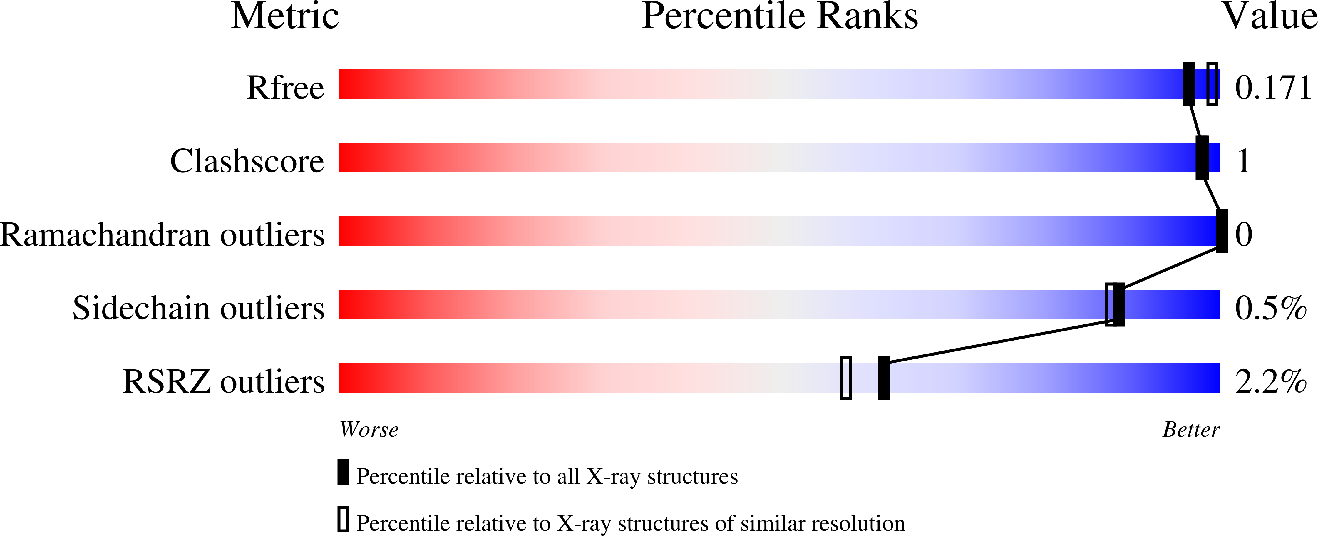
Experimental Data Snapshot
Entity ID: 1 | |||||
|---|---|---|---|---|---|
| Molecule | Chains | Sequence Length | Organism | Details | Image |
| Enoyl-[acyl-carrier-protein] reductase [NADH] | 277 | Mycobacterium kansasii | Mutation(s): 0 Gene Names: inhA, BZL29_3124, BZL30_8369 EC: 1.3.1.9 | 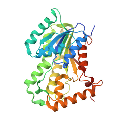 | |
UniProt | |||||
Find proteins for A0A1V3XDT0 (Mycobacterium kansasii) Explore A0A1V3XDT0 Go to UniProtKB: A0A1V3XDT0 | |||||
Entity Groups | |||||
| Sequence Clusters | 30% Identity50% Identity70% Identity90% Identity95% Identity100% Identity | ||||
| UniProt Group | A0A1V3XDT0 | ||||
Sequence AnnotationsExpand | |||||
| |||||
| Ligands 4 Unique | |||||
|---|---|---|---|---|---|
| ID | Chains | Name / Formula / InChI Key | 2D Diagram | 3D Interactions | |
| NAD (Subject of Investigation/LOI) Query on NAD | B [auth A] | NICOTINAMIDE-ADENINE-DINUCLEOTIDE C21 H27 N7 O14 P2 BAWFJGJZGIEFAR-NNYOXOHSSA-N |  | ||
| PO4 Query on PO4 | E [auth A] | PHOSPHATE ION O4 P NBIIXXVUZAFLBC-UHFFFAOYSA-K |  | ||
| EDO Query on EDO | D [auth A] | 1,2-ETHANEDIOL C2 H6 O2 LYCAIKOWRPUZTN-UHFFFAOYSA-N |  | ||
| K Query on K | C [auth A] | POTASSIUM ION K NPYPAHLBTDXSSS-UHFFFAOYSA-N |  | ||
| Length ( Å ) | Angle ( ˚ ) |
|---|---|
| a = 97.67 | α = 90 |
| b = 97.67 | β = 90 |
| c = 141.03 | γ = 120 |
| Software Name | Purpose |
|---|---|
| XDS | data reduction |
| XSCALE | data scaling |
| PHENIX | refinement |
| PDB_EXTRACT | data extraction |
| PHASER | phasing |
| PHENIX | model building |
| Coot | model building |
RCSB PDB (citation) is hosted by
RCSB PDB is a member of the

