7KSA

Crystal structure of human CYP3A4 with the caged inhibitor


Experimental Data Snapshot

  • Method: X-RAY DIFFRACTION
  • Resolution: 2.50 Å
  • R-Value Free: 0.266 
  • R-Value Work: 0.228 
  • R-Value Observed: 0.230 

wwPDB Validation   3D Report Full Report


Ligand Structure Quality Assessment 


This is version 1.2 of the entry. See complete history


Literature

Photosensitive Ru(II) Complexes as Inhibitors of the Major Human Drug Metabolizing Enzyme CYP3A4.

Toupin, N.Steinke, S.J.Nadella, S.Li, A.Rohrabaugh Jr., T.N.Samuels, E.R.Turro, C.Sevrioukova, I.F.Kodanko, J.J.

(2021) J Am Chem Soc 143: 9191-9205

  • DOI: https://doi.org/10.1021/jacs.1c04155
  • Primary Citation of Related Structures:  
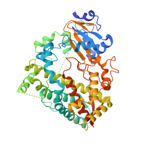
    7KS8, 7KSA

  • PubMed Abstract: 

    We report the synthesis and photochemical and biological characterization of the first selective and potent metal-based inhibitors of cytochrome P450 3A4 (CYP3A4), the major human drug metabolizing enzyme. Five Ru(II)-based derivatives were prepared from two analogs of the CYP3A4 inhibitor ritonavir, 4 and 6 : [Ru(tpy)(L)( 6 )]Cl 2 (tpy = 2,2':6',2″-terpyridine) with L = 6,6'-dimethyl-2,2'-bipyridine (Me 2 bpy; 8 ), dimethylbenzo[ i ]dipyrido[3,2- a :2',3'- c ]phenazine (Me 2 dppn; 10 ) and 3,6-dimethyl-10,15-diphenylbenzo[ i ]dipyrido[3,2- a :2',3'- c ]phenazine (Me 2 Ph 2 dppn; 11 ), [Ru(tpy)(Me 2 bpy)( 4 )]Cl 2 ( 7 ) and [Ru(tpy)(Me 2 dppn)( 4 )]Cl 2 ( 9 ). Photochemical release of 4 or 6 from 7 - 11 was demonstrated, and the spectrophotometric evaluation of 7 showed that it behaves similarly to free 4 (type II heme ligation) after irradiation with visible light but not in the dark. Unexpectedly, the intact Ru(II) complexes 7 and 8 were found to inhibit CYP3A4 potently and specifically through direct binding to the active site without heme ligation. Caged inhibitors 9 - 11 showed dual action properties by combining photoactivated dissociation of 4 or 6 with efficient 1 O 2 production. In prostate adenocarcinoma DU-145 cells, compound 9 had the best synergistic effect with vinblastine, the anticancer drug primarily metabolized by CYP3A4 in vivo . Thus, our study establishes a new paradigm in CYP inhibition using metalated complexes and suggests possible utilization of photoactive CYP3A4 inhibitory compounds in clinical applications, such as enhancement of therapeutic efficacy of anticancer drugs.


  • Organizational Affiliation

    Department of Chemistry, Wayne State University, 5101 Cass Avenue, Detroit, Michigan 48202, United States.


Macromolecules
Find similar proteins by:  (by identity cutoff)  |  3D Structure
Entity ID: 1
MoleculeChains Sequence LengthOrganismDetailsImage
Cytochrome P450 3A4487Homo sapiensMutation(s): 0 
Gene Names: CYP3A4CYP3A3
EC: 1.14.14.1 (PDB Primary Data), 1.14.14.56 (PDB Primary Data), 1.14.14.73 (PDB Primary Data), 1.14.14.55 (PDB Primary Data)
Membrane Entity: Yes 
UniProt & NIH Common Fund Data Resources
Find proteins for P08684 (Homo sapiens)
Explore P08684 
Go to UniProtKB:  P08684
PHAROS:  P08684
GTEx:  ENSG00000160868 
Entity Groups  
Sequence Clusters30% Identity50% Identity70% Identity90% Identity95% Identity100% Identity
UniProt GroupP08684
Sequence Annotations
Expand
  • Reference Sequence
Small Molecules
Ligands 2 Unique
IDChains Name / Formula / InChI Key2D Diagram3D Interactions
X8P (Subject of Investigation/LOI)
Query on X8P

Download Ideal Coordinates CCD File 
C [auth A](tert-butyl {1-[(1-oxo-3-phenyl-1-{[3-(pyridin-3-yl-kappaN)prop-1-en-1-yl]amino}propan-2-yl)sulfanyl]-3-phenylpropan-2-yl}carbamate)(6,6'-dimethyl-2,2'-bipyridine-kappa~2~N~1~,N~1'~)(1~2~,2~2~:2~6~,3~2~-terpyridine-kappa~3~N~1^{1~},N~2^{1~},N~3^{1~})ruthenium
C58 H62 N8 O3 Ru S
FVGJXMAFINABIT-VKKDUPNPSA-N
HEM
Query on HEM

Download Ideal Coordinates CCD File 
B [auth A]PROTOPORPHYRIN IX CONTAINING FE
C34 H32 Fe N4 O4
KABFMIBPWCXCRK-RGGAHWMASA-L
Experimental Data & Validation

Experimental Data

  • Method: X-RAY DIFFRACTION
  • Resolution: 2.50 Å
  • R-Value Free: 0.266 
  • R-Value Work: 0.228 
  • R-Value Observed: 0.230 
  • Space Group: I 2 2 2
Unit Cell:
Length ( Å )Angle ( ˚ )
a = 74.57α = 90
b = 95β = 90
c = 121.69γ = 90
Software Package:
Software NamePurpose
PHENIXrefinement
MOSFLMdata reduction
SCALAdata scaling
PHASERphasing

Structure Validation

View Full Validation Report


Ligand Structure Quality Assessment 


Entry History & Funding Information

Deposition Data


Funding OrganizationLocationGrant Number
National Institutes of Health/National Institute of Environmental Health Sciences (NIH/NIEHS)United StatesES025767

Revision History  (Full details and data files)

  • Version 1.0: 2021-06-02
    Type: Initial release
  • Version 1.1: 2021-07-28
    Changes: Database references
  • Version 1.2: 2023-10-18
    Changes: Data collection, Database references, Refinement description