7KS0

GluK2/K5 with 6-Cyano-7-nitroquinoxaline-2,3-dione (CNQX)


Experimental Data Snapshot

  • Method: ELECTRON MICROSCOPY
  • Resolution: 5.30 Å
  • Aggregation State: PARTICLE 
  • Reconstruction Method: SINGLE PARTICLE 

wwPDB Validation   3D Report Full Report


This is version 1.2 of the entry. See complete history


Literature

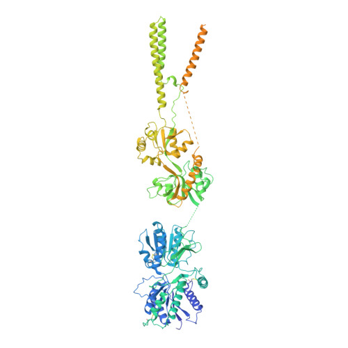
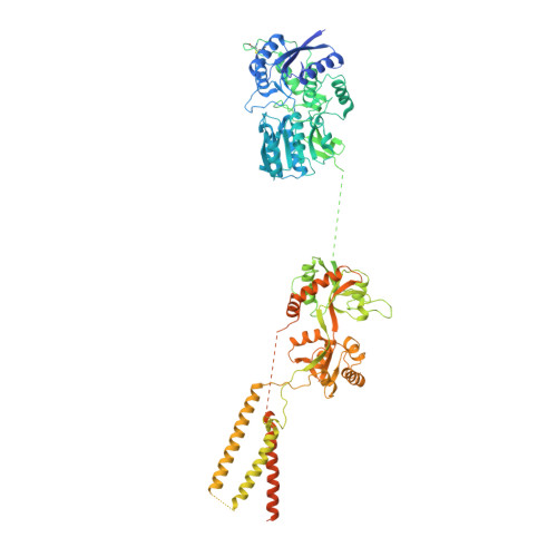
Architecture and structural dynamics of the heteromeric GluK2/K5 kainate receptor.

Khanra, N.Brown, P.M.Perozzo, A.M.Bowie, D.Meyerson, J.

(2021) Elife 10

  • DOI: https://doi.org/10.7554/eLife.66097
  • Primary Citation of Related Structures:  
    7KS0, 7KS3

  • PubMed Abstract: 

    Kainate receptors (KARs) are L-glutamate-gated ion channels that regulate synaptic transmission and modulate neuronal circuits. KARs have strict assembly rules and primarily function as heteromeric receptors in the brain. A longstanding question is how KAR heteromer subunits organize and coordinate together to fulfill their signature physiological roles. Here we report structures of the GluK2/GluK5 heteromer in apo, antagonist-bound, and desensitized states. The receptor assembles with two copies of each subunit, ligand binding domains arranged as two heterodimers and GluK5 subunits proximal to the channel. Strikingly, during desensitization, GluK2, but not GluK5, subunits undergo major structural rearrangements to facilitate channel closure. We show how the large conformational differences between antagonist-bound and desensitized states are mediated by the linkers connecting the pore helices to the ligand binding domains. This work presents the first KAR heteromer structure, reveals how its subunits are organized, and resolves how the heteromer can accommodate functionally distinct closed channel structures.


  • Organizational Affiliation

    Department of Physiology and Biophysics, Weill Cornell Medical College, New York, United States.


Macromolecules
Find similar proteins by:  (by identity cutoff)  |  3D Structure
Entity ID: 1
MoleculeChains Sequence LengthOrganismDetailsImage
Glutamate receptor ionotropic, kainate 5,Green fluorescent protein chimera
A, C
1,101Rattus norvegicusAequorea victoria
This entity is chimeric
Mutation(s): 0 
Gene Names: Grik5GFP
Membrane Entity: Yes 
UniProt
Find proteins for P42212 (Aequorea victoria)
Explore P42212 
Go to UniProtKB:  P42212
Find proteins for Q63273 (Rattus norvegicus)
Explore Q63273 
Go to UniProtKB:  Q63273
Entity Groups  
Sequence Clusters30% Identity50% Identity70% Identity90% Identity95% Identity100% Identity
UniProt GroupsP42212Q63273
Sequence Annotations
Expand
  • Reference Sequence
Find similar proteins by:  (by identity cutoff)  |  3D Structure
Entity ID: 2
MoleculeChains Sequence LengthOrganismDetailsImage
Glutamate receptor ionotropic, kainate 2
B, D
942Rattus norvegicusMutation(s): 0 
Gene Names: Grik2Glur6
Membrane Entity: Yes 
UniProt
Find proteins for P42260 (Rattus norvegicus)
Explore P42260 
Go to UniProtKB:  P42260
Entity Groups  
Sequence Clusters30% Identity50% Identity70% Identity90% Identity95% Identity100% Identity
UniProt GroupP42260
Sequence Annotations
Expand
  • Reference Sequence
Experimental Data & Validation

Experimental Data

  • Method: ELECTRON MICROSCOPY
  • Resolution: 5.30 Å
  • Aggregation State: PARTICLE 
  • Reconstruction Method: SINGLE PARTICLE 

Structure Validation

View Full Validation Report


Entry History 

Deposition Data

Revision History  (Full details and data files)

  • Version 1.0: 2021-03-24
    Type: Initial release
  • Version 1.1: 2021-03-31
    Changes: Database references
  • Version 1.2: 2021-07-07
    Changes: Derived calculations