7KL5

Structure of Calmodulin Bound to the Cardiac Ryanodine Receptor (RyR2) at Residues: Phe4246 to Val4271


Experimental Data Snapshot

  • Method: X-RAY DIFFRACTION
  • Resolution: 1.65 Å
  • R-Value Free: 0.245 
  • R-Value Work: 0.209 

wwPDB Validation   3D Report Full Report


This is version 1.2 of the entry. See complete history


Literature

The Crystal Structure of Calmodulin Bound to the Cardiac Ryanodine Receptor (RyR2) at Residues Phe4246-Val4271 Reveals a Fifth Calcium Binding Site.

Yu, Q.Anderson, D.E.Kaur, R.Fisher, A.J.Ames, J.B.

(2021) Biochemistry 60: 1088-1096

  • DOI: https://doi.org/10.1021/acs.biochem.1c00152
  • Primary Citation of Related Structures:  
    7KL5

  • PubMed Abstract: 

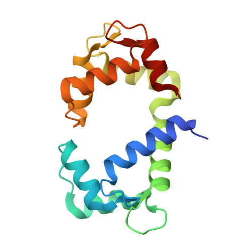
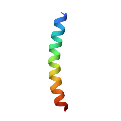
    Calmodulin (CaM) regulates the activity of a Ca 2+ channel known as the cardiac ryanodine receptor (RyR2), which facilitates the release of Ca 2+ from the sarcoplasmic reticulum during excitation-contraction coupling in cardiomyocytes. Mutations that disrupt this CaM-dependent channel inactivation result in cardiac arrhythmias. RyR2 contains three different CaM binding sites: CaMBD1 (residues 1940-1965), CaMBD2 (residues 3580-3611), and CaMBD3 (residues 4246-4275). Here, we report a crystal structure of Ca 2+ -bound CaM bound to RyR2 CaMBD3. The structure reveals Ca 2+ bound to the four EF-hands of CaM as well as a fifth Ca 2+ bound to CaM in the interdomain linker region involving Asp81 and Glu85. The CaM mutant E85A abolishes the binding of the fifth Ca 2+ and weakens the binding of CaMBD3 to Ca 2+ -bound CaM. Thus, the binding of the fifth Ca 2+ is important for stabilizing the complex in solution and is not a crystalline artifact. The CaMBD3 peptide in the complex adopts an α-helix (between Phe4246 and Val4271) that interacts with both lobes of CaM. Hydrophobic residues in the CaMBD3 helix (Leu4255 and Leu4259) form intermolecular contacts with the CaM N-lobe, and the CaMBD3 mutations (L4255A and L4259A) each weaken the binding of CaM to RyR2. Aromatic residues on the opposite side of the CaMBD3 helix (Phe4246 and Tyr4250) interact with the CaM C-lobe, but the mutants (F4246A and Y4250A) have no detectable effect on CaM binding in solution. We suggest that the binding of CaM to CaMBD3 and the binding of a fifth Ca 2+ to CaM may contribute to the regulation of RyR2 channel function.


  • Organizational Affiliation

    Department of Chemistry, University of California, Davis, California 95616, United States.


Macromolecules
Find similar proteins by:  (by identity cutoff)  |  3D Structure
Entity ID: 1
MoleculeChains Sequence LengthOrganismDetailsImage
Calmodulin-1149Homo sapiensMutation(s): 0 
Gene Names: CALM1CALMCAMCAM1
UniProt & NIH Common Fund Data Resources
Find proteins for P0DP23 (Homo sapiens)
Explore P0DP23 
Go to UniProtKB:  P0DP23
PHAROS:  P0DP23
GTEx:  ENSG00000198668 
Entity Groups  
Sequence Clusters30% Identity50% Identity70% Identity90% Identity95% Identity100% Identity
UniProt GroupP0DP23
Sequence Annotations
Expand
  • Reference Sequence
Find similar proteins by:  (by identity cutoff)  |  3D Structure
Entity ID: 2
MoleculeChains Sequence LengthOrganismDetailsImage
Ryanodine receptor 230Homo sapiensMutation(s): 0 
Gene Names: RYR2
UniProt & NIH Common Fund Data Resources
Find proteins for Q92736 (Homo sapiens)
Explore Q92736 
Go to UniProtKB:  Q92736
PHAROS:  Q92736
GTEx:  ENSG00000198626 
Entity Groups  
Sequence Clusters30% Identity50% Identity70% Identity90% Identity95% Identity100% Identity
UniProt GroupQ92736
Sequence Annotations
Expand
  • Reference Sequence
Experimental Data & Validation

Experimental Data

  • Method: X-RAY DIFFRACTION
  • Resolution: 1.65 Å
  • R-Value Free: 0.245 
  • R-Value Work: 0.209 
  • Space Group: P 1 21 1
Unit Cell:
Length ( Å )Angle ( ˚ )
a = 33.169α = 90
b = 62.403β = 107.494
c = 43.19γ = 90
Software Package:
Software NamePurpose
REFMACrefinement
XDSdata reduction
Aimlessdata scaling
PHASERphasing

Structure Validation

View Full Validation Report


Entry History & Funding Information

Deposition Data


Funding OrganizationLocationGrant Number
National Institutes of Health/National Eye Institute (NIH/NEI)United StatesR01 EY012347

Revision History  (Full details and data files)

  • Version 1.0: 2021-04-07
    Type: Initial release
  • Version 1.1: 2021-04-21
    Changes: Database references
  • Version 1.2: 2023-10-18
    Changes: Data collection, Database references, Refinement description