7KHF

CryoEM structure of LILRB1 D3D4 domain-inserted antibody MDB1 Fab in complex with Plasmodium RIFIN (PF3D7_1373400) V2 domain


Experimental Data Snapshot

  • Method: ELECTRON MICROSCOPY
  • Resolution: 3.54 Å
  • Aggregation State: PARTICLE 
  • Reconstruction Method: SINGLE PARTICLE 

wwPDB Validation   3D Report Full Report


This is version 1.2 of the entry. See complete history


Literature

Structural basis of malaria RIFIN binding by LILRB1-containing antibodies.

Chen, Y.Xu, K.Piccoli, L.Foglierini, M.Tan, J.Jin, W.Gorman, J.Tsybovsky, Y.Zhang, B.Traore, B.Silacci-Fregni, C.Daubenberger, C.Crompton, P.D.Geiger, R.Sallusto, F.Kwong, P.D.Lanzavecchia, A.

(2021) Nature 592: 639-643

  • DOI: https://doi.org/10.1038/s41586-021-03378-6
  • Primary Citation of Related Structures:  
    7KFK, 7KHF

  • PubMed Abstract: 

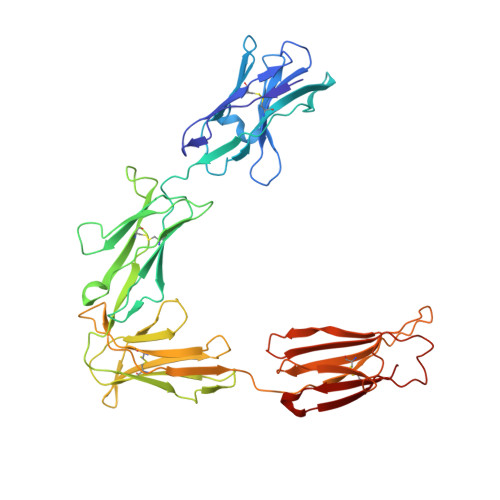
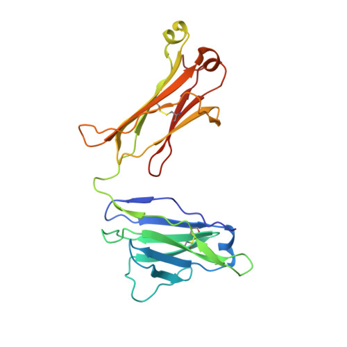
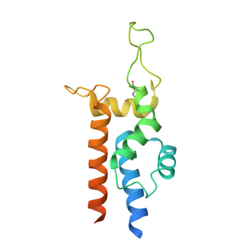
    Some Plasmodium falciparum repetitive interspersed families of polypeptides (RIFINs)-variant surface antigens that are expressed on infected erythrocytes 1 -bind to the inhibitory receptor LAIR1, and insertion of DNA that encodes LAIR1 into immunoglobulin genes generates RIFIN-specific antibodies 2,3 . Here we address the general relevance of this finding by searching for antibodies that incorporate LILRB1, another inhibitory receptor that binds to β2 microglobulin and RIFINs through their apical domains 4,5 . By screening plasma from a cohort of donors from Mali, we identified individuals with LILRB1-containing antibodies. B cell clones isolated from three donors showed large DNA insertions in the switch region that encodes non-apical LILRB1 extracellular domain 3 and 4 (D3D4) or D3 alone in the variable-constant (VH-CH1) elbow. Through mass spectrometry and binding assays, we identified a large set of RIFINs that bind to LILRB1 D3. Crystal and cryo-electron microscopy structures of a RIFIN in complex with either LILRB1 D3D4 or a D3D4-containing antibody Fab revealed a mode of RIFIN-LILRB1 D3 interaction that is similar to that of RIFIN-LAIR1. The Fab showed an unconventional triangular architecture with the inserted LILRB1 domains opening up the VH-CH1 elbow without affecting VH-VL or CH1-CL pairing. Collectively, these findings show that RIFINs bind to LILRB1 through D3 and illustrate, with a naturally selected example, the general principle of creating novel antibodies by inserting receptor domains into the VH-CH1 elbow.


  • Organizational Affiliation

    Institute for Research in Biomedicine, Università della Svizzera italiana, Bellinzona, Switzerland.


Macromolecules
Find similar proteins by:  (by identity cutoff)  |  3D Structure
Entity ID: 1
MoleculeChains Sequence LengthOrganismDetailsImage
MDB1 Fab heavy chainA [auth H],
C [auth A]
427Homo sapiensMutation(s): 0 
Entity Groups  
Sequence Clusters30% Identity50% Identity70% Identity90% Identity95% Identity100% Identity
Sequence Annotations
Expand
  • Reference Sequence
Find similar proteins by:  (by identity cutoff)  |  3D Structure
Entity ID: 2
MoleculeChains Sequence LengthOrganismDetailsImage
MDB1 Fab light chainB [auth L],
D [auth B]
214Homo sapiensMutation(s): 0 
Entity Groups  
Sequence Clusters30% Identity50% Identity70% Identity90% Identity95% Identity100% Identity
Sequence Annotations
Expand
  • Reference Sequence
Find similar proteins by:  (by identity cutoff)  |  3D Structure
Entity ID: 3
MoleculeChains Sequence LengthOrganismDetailsImage
RifinE [auth D],
F [auth C]
151Plasmodium falciparum 3D7Mutation(s): 0 
Gene Names: PF3D7_1373400
UniProt
Find proteins for C0H5N9 (Plasmodium falciparum (isolate 3D7))
Explore C0H5N9 
Go to UniProtKB:  C0H5N9
Entity Groups  
Sequence Clusters30% Identity50% Identity70% Identity90% Identity95% Identity100% Identity
UniProt GroupC0H5N9
Sequence Annotations
Expand
  • Reference Sequence
Oligosaccharides

Help

Entity ID: 4
MoleculeChains Length2D Diagram Glycosylation3D Interactions
2-acetamido-2-deoxy-beta-D-glucopyranose-(1-4)-2-acetamido-2-deoxy-beta-D-glucopyranoseG [auth E],
H [auth F],
I [auth G],
J [auth I]
2N-Glycosylation
Glycosylation Resources
GlyTouCan:  G42666HT
GlyCosmos:  G42666HT
GlyGen:  G42666HT
Small Molecules
Ligands 1 Unique
IDChains Name / Formula / InChI Key2D Diagram3D Interactions
NAG
Query on NAG

Download Ideal Coordinates CCD File 
K [auth H],
L [auth A]
2-acetamido-2-deoxy-beta-D-glucopyranose
C8 H15 N O6
OVRNDRQMDRJTHS-FMDGEEDCSA-N
Experimental Data & Validation

Experimental Data

  • Method: ELECTRON MICROSCOPY
  • Resolution: 3.54 Å
  • Aggregation State: PARTICLE 
  • Reconstruction Method: SINGLE PARTICLE 
EM Software:
TaskSoftware PackageVersion
RECONSTRUCTIONcryoSPARC
MODEL REFINEMENTPHENIX

Structure Validation

View Full Validation Report


Entry History & Funding Information

Deposition Data


Funding OrganizationLocationGrant Number
Other privateUnited StatesSimons Foundation SF349247
National Institutes of Health/National Institute of General Medical Sciences (NIH/NIGMS)United StatesU24 GM129539

Revision History  (Full details and data files)

  • Version 1.0: 2021-03-31
    Type: Initial release
  • Version 1.1: 2021-04-14
    Changes: Database references
  • Version 1.2: 2021-05-05
    Changes: Database references