7KD4

Structure of the C-terminal domain of the Menangle virus phosphoprotein (residues 329 -388), fused to MBP. Space group P21.


Experimental Data Snapshot

  • Method: X-RAY DIFFRACTION
  • Resolution: 1.31 Å
  • R-Value Free: 0.196 
  • R-Value Work: 0.150 

wwPDB Validation   3D Report Full Report


This is version 1.2 of the entry. See complete history


Literature

Structural Analysis of the Menangle Virus P Protein Reveals a Soft Boundary between Ordered and Disordered Regions.

Webby, M.N.Herr, N.Bulloch, E.M.M.Schmitz, M.Keown, J.R.Goldstone, D.C.Kingston, R.L.

(2021) Viruses 13

  • DOI: https://doi.org/10.3390/v13091737
  • Primary Citation of Related Structures:  
    7KD4, 7KD5

  • PubMed Abstract: 

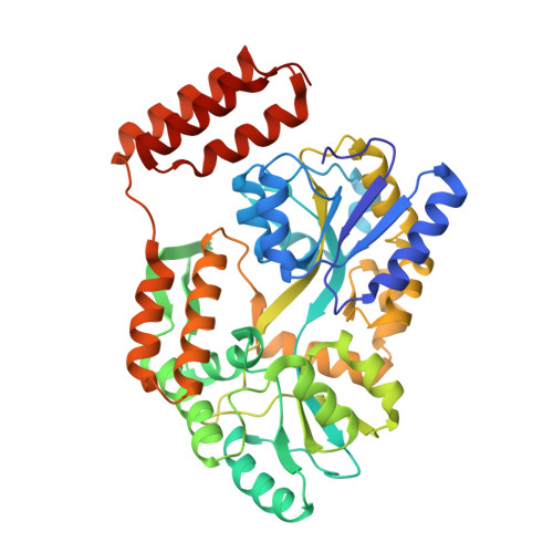
    The paramyxoviral phosphoprotein (P protein) is the non-catalytic subunit of the viral RNA polymerase, and coordinates many of the molecular interactions required for RNA synthesis. All paramyxoviral P proteins oligomerize via a centrally located coiled-coil that is connected to a downstream binding domain by a dynamic linker. The C-terminal region of the P protein coordinates interactions between the catalytic subunit of the polymerase, and the viral nucleocapsid housing the genomic RNA. The inherent flexibility of the linker is believed to facilitate polymerase translocation. Here we report biophysical and structural characterization of the C-terminal region of the P protein from Menangle virus (MenV), a bat-borne paramyxovirus with zoonotic potential. The MenV P protein is tetrameric but can dissociate into dimers at sub-micromolar protein concentrations. The linker is globally disordered and can be modeled effectively as a worm-like chain. However, NMR analysis suggests very weak local preferences for alpha-helical and extended beta conformation exist within the linker. At the interface between the disordered linker and the structured C-terminal binding domain, a gradual disorder-to-order transition occurs, with X-ray crystallographic analysis revealing a dynamic interfacial structure that wraps the surface of the binding domain.


  • Organizational Affiliation

    School of Biological Sciences, University of Auckland, Auckland 1010, New Zealand.


Macromolecules
Find similar proteins by:  (by identity cutoff)  |  3D Structure
Entity ID: 1
MoleculeChains Sequence LengthOrganismDetailsImage
Maltodextrin-binding protein and Phosphoprotein fusion protein
A, B
431Serratia sp. FS14Menangle virus
This entity is chimeric
Mutation(s): 1 
Gene Names: malEJW3994V/P
UniProt
Find proteins for P0AEX9 (Escherichia coli (strain K12))
Explore P0AEX9 
Go to UniProtKB:  P0AEX9
Find proteins for Q91MK1 (Menangle virus)
Explore Q91MK1 
Go to UniProtKB:  Q91MK1
Entity Groups  
Sequence Clusters30% Identity50% Identity70% Identity90% Identity95% Identity100% Identity
UniProt GroupsQ91MK1P0AEX9
Sequence Annotations
Expand
  • Reference Sequence
Oligosaccharides

Help

Entity ID: 2
MoleculeChains Length2D Diagram Glycosylation3D Interactions
alpha-D-glucopyranose-(1-4)-alpha-D-glucopyranose
C, D
2N/A
Glycosylation Resources
GlyTouCan:  G07411ON
GlyCosmos:  G07411ON
Small Molecules
Ligands 1 Unique
IDChains Name / Formula / InChI Key2D Diagram3D Interactions
SO4
Query on SO4

Download Ideal Coordinates CCD File 
E [auth A]
F [auth A]
G [auth A]
H [auth A]
I [auth A]
E [auth A],
F [auth A],
G [auth A],
H [auth A],
I [auth A],
J [auth A],
K [auth A],
L [auth A],
M [auth A],
N [auth B],
O [auth B],
P [auth B],
Q [auth B],
R [auth B],
S [auth B],
T [auth B]
SULFATE ION
O4 S
QAOWNCQODCNURD-UHFFFAOYSA-L
Biologically Interesting Molecules (External Reference) 1 Unique
Experimental Data & Validation

Experimental Data

  • Method: X-RAY DIFFRACTION
  • Resolution: 1.31 Å
  • R-Value Free: 0.196 
  • R-Value Work: 0.150 
  • Space Group: P 1 21 1
Unit Cell:
Length ( Å )Angle ( ˚ )
a = 57.561α = 90
b = 70.074β = 96.472
c = 111.739γ = 90
Software Package:
Software NamePurpose
REFMACrefinement
HKL-2000data reduction
SCALEPACKdata scaling
PHASERphasing
Cootmodel building

Structure Validation

View Full Validation Report


Entry History & Funding Information

Deposition Data


Funding OrganizationLocationGrant Number
Marsden FundNew ZealandUOA1202

Revision History  (Full details and data files)

  • Version 1.0: 2021-09-15
    Type: Initial release
  • Version 1.1: 2021-10-13
    Changes: Database references
  • Version 1.2: 2023-10-18
    Changes: Data collection, Refinement description