7KCH

Myosin XI-F-actin complex


Experimental Data Snapshot

  • Method: ELECTRON MICROSCOPY
  • Resolution: 4.33 Å
  • Aggregation State: FILAMENT 
  • Reconstruction Method: HELICAL 

wwPDB Validation   3D Report Full Report


This is version 1.4 of the entry. See complete history


Literature

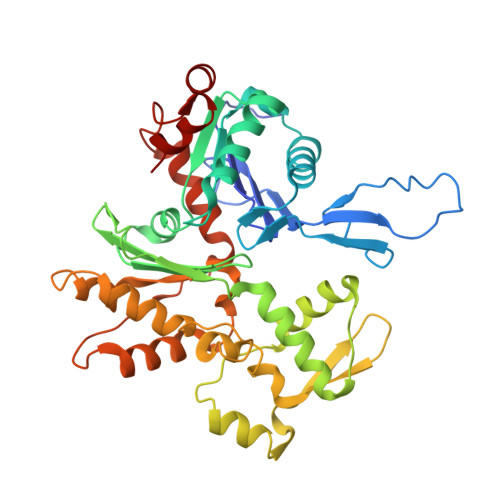
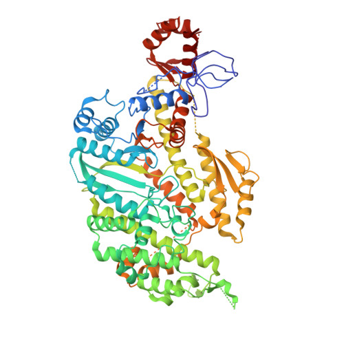
Optical control of fast and processive engineered myosins in vitro and in living cells.

Ruijgrok, P.V.Ghosh, R.P.Zemsky, S.Nakamura, M.Gong, R.Ning, L.Chen, R.Vachharajani, V.T.Chu, A.E.Anand, N.Eguchi, R.R.Huang, P.S.Lin, M.Z.Alushin, G.M.Liphardt, J.T.Bryant, Z.

(2021) Nat Chem Biol 17: 540-548

  • DOI: https://doi.org/10.1038/s41589-021-00740-7
  • Primary Citation of Related Structures:  
    7KCH

  • PubMed Abstract: 

    Precision tools for spatiotemporal control of cytoskeletal motor function are needed to dissect fundamental biological processes ranging from intracellular transport to cell migration and division. Direct optical control of motor speed and direction is one promising approach, but it remains a challenge to engineer controllable motors with desirable properties such as the speed and processivity required for transport applications in living cells. Here, we develop engineered myosin motors that combine large optical modulation depths with high velocities, and create processive myosin motors with optically controllable directionality. We characterize the performance of the motors using in vitro motility assays, single-molecule tracking and live-cell imaging. Bidirectional processive motors move efficiently toward the tips of cellular protrusions in the presence of blue light, and can transport molecular cargo in cells. Robust gearshifting myosins will further enable programmable transport in contexts ranging from in vitro active matter reconstitutions to microfabricated systems that harness molecular propulsion.


  • Organizational Affiliation

    Department of Bioengineering, Stanford University, Stanford, CA, USA.


Macromolecules
Find similar proteins by:  (by identity cutoff)  |  3D Structure
Entity ID: 1
MoleculeChains Sequence LengthOrganismDetailsImage
Actin, alpha skeletal muscleA [auth G],
C [auth B],
D [auth C]
377Gallus gallusMutation(s): 0 
Gene Names: ACTA1ACTA
UniProt
Find proteins for P68139 (Gallus gallus)
Explore P68139 
Go to UniProtKB:  P68139
Entity Groups  
Sequence Clusters30% Identity50% Identity70% Identity90% Identity95% Identity100% Identity
UniProt GroupP68139
Sequence Annotations
Expand
  • Reference Sequence
Find similar proteins by:  (by identity cutoff)  |  3D Structure
Entity ID: 2
MoleculeChains Sequence LengthOrganismDetailsImage
Unconventional myosin heavy chainB [auth A]735Chara corallinaMutation(s): 0 
Gene Names: ccm
UniProt
Find proteins for Q9SSU1 (Chara corallina)
Explore Q9SSU1 
Go to UniProtKB:  Q9SSU1
Entity Groups  
Sequence Clusters30% Identity50% Identity70% Identity90% Identity95% Identity100% Identity
UniProt GroupQ9SSU1
Sequence Annotations
Expand
  • Reference Sequence
Experimental Data & Validation

Experimental Data

  • Method: ELECTRON MICROSCOPY
  • Resolution: 4.33 Å
  • Aggregation State: FILAMENT 
  • Reconstruction Method: HELICAL 
EM Software:
TaskSoftware PackageVersion
RECONSTRUCTIONRELION3.0

Structure Validation

View Full Validation Report


Entry History & Funding Information

Deposition Data


Funding OrganizationLocationGrant Number
National Institutes of Health/National Institute of General Medical Sciences (NIH/NIGMS)United StatesR01-GM114627

Revision History  (Full details and data files)

  • Version 1.0: 2021-01-13
    Type: Initial release
  • Version 1.1: 2021-02-17
    Changes: Database references
  • Version 1.2: 2021-03-10
    Changes: Database references
  • Version 1.3: 2021-05-05
    Changes: Database references
  • Version 1.4: 2024-03-06
    Changes: Data collection, Database references