7K81

KIR3DL1*005 in complex with HLA-A*24:02 presenting the RYPLTFGW peptide


Experimental Data Snapshot

  • Method: X-RAY DIFFRACTION
  • Resolution: 2.00 Å
  • R-Value Free: 0.243 
  • R-Value Work: 0.190 
  • R-Value Observed: 0.193 

wwPDB Validation   3D Report Full Report


Ligand Structure Quality Assessment 


This is version 1.3 of the entry. See complete history


Literature

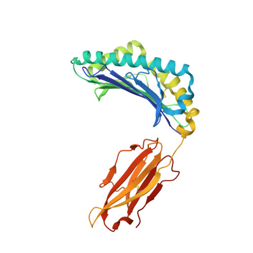
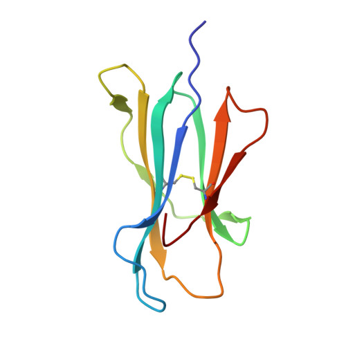
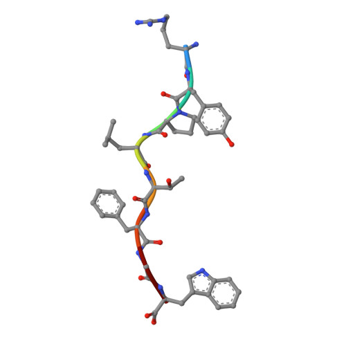
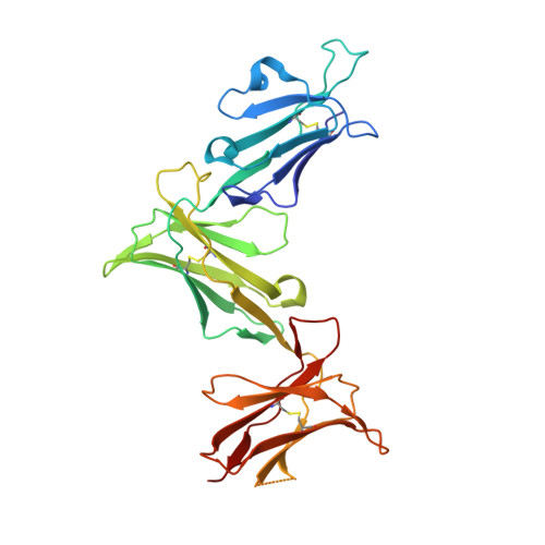
The Role of the HLA Class I alpha 2 Helix in Determining Ligand Hierarchy for the Killer Cell Ig-like Receptor 3DL1.

Saunders, P.M.MacLachlan, B.J.Widjaja, J.Wong, S.C.Oates, C.V.L.Rossjohn, J.Vivian, J.P.Brooks, A.G.

(2021) J Immunol 206: 849-860

  • DOI: https://doi.org/10.4049/jimmunol.2001109
  • Primary Citation of Related Structures:  
    7K80, 7K81

  • PubMed Abstract: 

    HLA class I molecules that represent ligands for the inhibitory killer cell Ig-like receptor (KIR) 3DL1 found on NK cells are categorically defined as those HLA-A and HLA-B allotypes containing the Bw4 motif, yet KIR3DL1 demonstrates hierarchical recognition of these HLA-Bw4 ligands. To better understand the molecular basis underpinning differential KIR3DL1 recognition, the HLA-A Bw4 family of allotypes were investigated. Transfected human 721.221 cells expressing HLA-A*32:01 strongly inhibited primary human KIR3DL1 + NK cells, whereas HLA-A*24:02 and HLA-A*23:01 displayed intermediate potency and HLA-A*25:01 failed to inhibit activation of KIR3DL1 + NK cells. Structural studies demonstrated that recognition of HLA-A*24:02 by KIR3DL1 used identical contacts as the potent HLA-B*57:01 ligand. Namely, the D1-D2 domains of KIR3DL1 were placed over the α1 helix and α2 helix of the HLA-A*24:02 binding cleft, respectively, whereas the D0 domain contacted the side of the HLA-A*24:02 molecule. Nevertheless, functional analyses showed KIR3DL1 recognition of HLA-A*24:02 was more sensitive to substitutions within the α2 helix of HLA-A*24:02, including residues Ile 142 and Lys 144 Furthermore, the presence of Thr 149 in the α2 helix of HLA-A*25:01 abrogated KIR3DL1 + NK inhibition. Together, these data demonstrate a role for the HLA class I α2 helix in determining the hierarchy of KIR3DL1 ligands. Thus, recognition of HLA class I is dependent on a complex interplay between the peptide repertoire, polymorphisms within and proximal to the Bw4 motif, and the α2 helix. Collectively, the data furthers our understanding of KIR3DL1 ligands and will inform genetic association and immunogenetics studies examining the role of KIR3DL1 in disease settings.


  • Organizational Affiliation

    Department of Microbiology and Immunology, Peter Doherty Institute for Infection and Immunity, The University of Melbourne, Melbourne, Victoria 3000, Australia; philippa.saunders@unimelb.edu.au agbrooks@unimelb.edu.au.


Macromolecules
Find similar proteins by:  (by identity cutoff)  |  3D Structure
Entity ID: 1
MoleculeChains Sequence LengthOrganismDetailsImage
MHC class I antigen276Homo sapiensMutation(s): 0 
Gene Names: HLA-A
UniProt
Find proteins for A0A5H2UYS3 (Homo sapiens)
Explore A0A5H2UYS3 
Go to UniProtKB:  A0A5H2UYS3
Entity Groups  
Sequence Clusters30% Identity50% Identity70% Identity90% Identity95% Identity100% Identity
UniProt GroupA0A5H2UYS3
Sequence Annotations
Expand
  • Reference Sequence
Find similar proteins by:  (by identity cutoff)  |  3D Structure
Entity ID: 2
MoleculeChains Sequence LengthOrganismDetailsImage
Beta-2-microglobulin100Homo sapiensMutation(s): 0 
Gene Names: B2MCDABP0092HDCMA22P
UniProt & NIH Common Fund Data Resources
Find proteins for P61769 (Homo sapiens)
Explore P61769 
Go to UniProtKB:  P61769
PHAROS:  P61769
GTEx:  ENSG00000166710 
Entity Groups  
Sequence Clusters30% Identity50% Identity70% Identity90% Identity95% Identity100% Identity
UniProt GroupP61769
Sequence Annotations
Expand
  • Reference Sequence

Find similar proteins by:  Sequence   |   3D Structure  

Entity ID: 3
MoleculeChains Sequence LengthOrganismDetailsImage
ARG-TYR-PRO-LEU-THR-PHE-GLY-TRP8synthetic constructMutation(s): 0 
UniProt
Find proteins for P04601 (Human immunodeficiency virus type 1 group M subtype B (isolate HXB2))
Explore P04601 
Go to UniProtKB:  P04601
Entity Groups  
UniProt GroupP04601
Sequence Annotations
Expand
  • Reference Sequence
Find similar proteins by:  (by identity cutoff)  |  3D Structure
Entity ID: 4
MoleculeChains Sequence LengthOrganismDetailsImage
KIR3DL1D [auth G]289Homo sapiensMutation(s): 0 
Gene Names: KIR3DL1
UniProt & NIH Common Fund Data Resources
Find proteins for P43629 (Homo sapiens)
Explore P43629 
Go to UniProtKB:  P43629
PHAROS:  P43629
GTEx:  ENSG00000167633 
Entity Groups  
Sequence Clusters30% Identity50% Identity70% Identity90% Identity95% Identity100% Identity
UniProt GroupP43629
Sequence Annotations
Expand
  • Reference Sequence
Experimental Data & Validation

Experimental Data

  • Method: X-RAY DIFFRACTION
  • Resolution: 2.00 Å
  • R-Value Free: 0.243 
  • R-Value Work: 0.190 
  • R-Value Observed: 0.193 
  • Space Group: P 1
Unit Cell:
Length ( Å )Angle ( ˚ )
a = 47.807α = 94.32
b = 61.385β = 99.82
c = 66.989γ = 105.97
Software Package:
Software NamePurpose
PHENIXrefinement
MOSFLMdata reduction
SCALAdata scaling
PHASERphasing

Structure Validation

View Full Validation Report


Ligand Structure Quality Assessment 


Entry History 

Deposition Data

Revision History  (Full details and data files)

  • Version 1.0: 2020-12-09
    Type: Initial release
  • Version 1.1: 2021-01-27
    Changes: Database references
  • Version 1.2: 2021-02-17
    Changes: Database references
  • Version 1.3: 2023-10-18
    Changes: Data collection, Database references, Refinement description