7K20

Cryo-EM structure of pyrene-labeled ADP-actin filaments

  • Classification: CYTOSOLIC PROTEIN
  • Organism(s): Gallus gallus
  • Mutation(s): No 

  • Deposited: 2020-09-08 Released: 2020-11-04 
  • Deposition Author(s): Chou, S.Z., Pollard, T.D.
  • Funding Organization(s): National Institutes of Health/National Institute of General Medical Sciences (NIH/NIGMS)

Experimental Data Snapshot

  • Method: ELECTRON MICROSCOPY
  • Resolution: 3.20 Å
  • Aggregation State: FILAMENT 
  • Reconstruction Method: HELICAL 

wwPDB Validation   3D Report Full Report


This is version 1.1 of the entry. See complete history


Literature

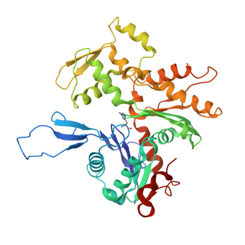
Cryo-electron microscopy structures of pyrene-labeled ADP-P i - and ADP-actin filaments.

Chou, S.Z.Pollard, T.D.

(2020) Nat Commun 11: 5897-5897

  • DOI: https://doi.org/10.1038/s41467-020-19762-1
  • Primary Citation of Related Structures:  
    7K20, 7K21

  • PubMed Abstract: 

    Since the fluorescent reagent N-(1-pyrene)iodoacetamide was first used to label skeletal muscle actin in 1981, the pyrene-labeled actin has become the most widely employed tool to measure the kinetics of actin polymerization and the interaction between actin and actin-binding proteins. Here we report high-resolution cryo-electron microscopy structures of actin filaments with N-1-pyrene conjugated to cysteine 374 and either ADP (3.2 Å) or ADP-phosphate (3.0 Å) in the active site. Polymerization buries pyrene in a hydrophobic cavity between subunits along the long-pitch helix with only minor differences in conformation compared with native actin filaments. These structures explain how polymerization increases the fluorescence 20-fold, how myosin and cofilin binding to filaments reduces the fluorescence, and how profilin binding to actin monomers increases the fluorescence.


  • Organizational Affiliation

    Department of Molecular Cellular and Developmental Biology, Yale University, PO Box 208103, New Haven, CT, 06520-8103, USA.


Macromolecules
Find similar proteins by:  (by identity cutoff)  |  3D Structure
Entity ID: 1
MoleculeChains Sequence LengthOrganismDetailsImage
Actin, alpha skeletal muscle
A, B, C, D
375Gallus gallusMutation(s): 0 
UniProt
Find proteins for P68139 (Gallus gallus)
Explore P68139 
Go to UniProtKB:  P68139
Entity Groups  
Sequence Clusters30% Identity50% Identity70% Identity90% Identity95% Identity100% Identity
UniProt GroupP68139
Sequence Annotations
Expand
  • Reference Sequence
Small Molecules
Ligands 3 Unique
IDChains Name / Formula / InChI Key2D Diagram3D Interactions
ADP (Subject of Investigation/LOI)
Query on ADP

Download Ideal Coordinates CCD File 
F [auth A],
I [auth B],
L [auth C],
O [auth D]
ADENOSINE-5'-DIPHOSPHATE
C10 H15 N5 O10 P2
XTWYTFMLZFPYCI-KQYNXXCUSA-N
1T4 (Subject of Investigation/LOI)
Query on 1T4

Download Ideal Coordinates CCD File 
G [auth A],
J [auth B],
M [auth C],
P [auth D]
N-(pyren-1-yl)acetamide
C18 H13 N O
GJNGMQYHOZFQNX-UHFFFAOYSA-N
MG
Query on MG

Download Ideal Coordinates CCD File 
E [auth A],
H [auth B],
K [auth C],
N [auth D]
MAGNESIUM ION
Mg
JLVVSXFLKOJNIY-UHFFFAOYSA-N
Modified Residues  1 Unique
IDChains TypeFormula2D DiagramParent
HIC
Query on HIC
A, B, C, D
L-PEPTIDE LINKINGC7 H11 N3 O2HIS
Experimental Data & Validation

Experimental Data

  • Method: ELECTRON MICROSCOPY
  • Resolution: 3.20 Å
  • Aggregation State: FILAMENT 
  • Reconstruction Method: HELICAL 
EM Software:
TaskSoftware PackageVersion
RECONSTRUCTIONRELION3.08
MODEL REFINEMENTPHENIX3.12

Structure Validation

View Full Validation Report


Entry History & Funding Information

Deposition Data


Funding OrganizationLocationGrant Number
National Institutes of Health/National Institute of General Medical Sciences (NIH/NIGMS)United StatesR01GM026338

Revision History  (Full details and data files)

  • Version 1.0: 2020-11-04
    Type: Initial release
  • Version 1.1: 2020-12-02
    Changes: Database references