7JY3

Structure of HbA with compound 23 (PF-07059013)


Experimental Data Snapshot

  • Method: X-RAY DIFFRACTION
  • Resolution: 1.48 Å
  • R-Value Free: 0.207 
  • R-Value Work: 0.180 
  • R-Value Observed: 0.181 

wwPDB Validation   3D Report Full Report


Ligand Structure Quality Assessment 


This is version 1.2 of the entry. See complete history


Literature

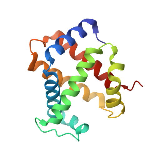
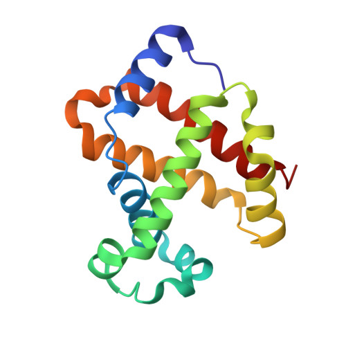
PF-07059013: A Noncovalent Modulator of Hemoglobin for Treatment of Sickle Cell Disease.

Gopalsamy, A.Aulabaugh, A.E.Barakat, A.Beaumont, K.C.Cabral, S.Canterbury, D.P.Casimiro-Garcia, A.Chang, J.S.Chen, M.Z.Choi, C.Dow, R.L.Fadeyi, O.O.Feng, X.France, S.P.Howard, R.M.Janz, J.M.Jasti, J.Jasuja, R.Jones, L.H.King-Ahmad, A.Knee, K.M.Kohrt, J.T.Limberakis, C.Liras, S.Martinez, C.A.McClure, K.F.Narayanan, A.Narula, J.Novak, J.J.O'Connell, T.N.Parikh, M.D.Piotrowski, D.W.Plotnikova, O.Robinson, R.P.Sahasrabudhe, P.V.Sharma, R.Thuma, B.A.Vasa, D.Wei, L.Wenzel, A.Z.Withka, J.M.Xiao, J.Yayla, H.G.

(2021) J Med Chem 64: 326-342

  • DOI: https://doi.org/10.1021/acs.jmedchem.0c01518
  • Primary Citation of Related Structures:  
    7JXZ, 7JY0, 7JY1, 7JY3

  • PubMed Abstract: 

    Sickle cell disease (SCD) is a genetic disorder caused by a single point mutation (β6 Glu → Val) on the β-chain of adult hemoglobin (HbA) that results in sickled hemoglobin (HbS). In the deoxygenated state, polymerization of HbS leads to sickling of red blood cells (RBC). Several downstream consequences of polymerization and RBC sickling include vaso-occlusion, hemolytic anemia, and stroke. We report the design of a noncovalent modulator of HbS, clinical candidate PF-07059013 ( 23 ). The seminal hit molecule was discovered by virtual screening and confirmed through a series of biochemical and biophysical studies. After a significant optimization effort, we arrived at 23 , a compound that specifically binds to Hb with nanomolar affinity and displays strong partitioning into RBCs. In a 2-week multiple dose study using Townes SCD mice, 23 showed a 37.8% (±9.0%) reduction in sickling compared to vehicle treated mice. 23 (PF-07059013) has advanced to phase 1 clinical trials.


  • Organizational Affiliation

    Pfizer Medicine Design, Pfizer Worldwide Research and Development, Cambridge, Massachusetts 02139, United States.


Macromolecules
Find similar proteins by:  (by identity cutoff)  |  3D Structure
Entity ID: 1
MoleculeChains Sequence LengthOrganismDetailsImage
Hemoglobin subunit alpha
A, C
141Homo sapiensMutation(s): 0 
Gene Names: HBA1HBA2
UniProt & NIH Common Fund Data Resources
Find proteins for P69905 (Homo sapiens)
Explore P69905 
Go to UniProtKB:  P69905
PHAROS:  P69905
Entity Groups  
Sequence Clusters30% Identity50% Identity70% Identity90% Identity95% Identity100% Identity
UniProt GroupP69905
Sequence Annotations
Expand
  • Reference Sequence
Find similar proteins by:  (by identity cutoff)  |  3D Structure
Entity ID: 2
MoleculeChains Sequence LengthOrganismDetailsImage
Hemoglobin subunit beta
B, D
146Homo sapiensMutation(s): 0 
Gene Names: HBB
UniProt & NIH Common Fund Data Resources
Find proteins for P68871 (Homo sapiens)
Explore P68871 
Go to UniProtKB:  P68871
PHAROS:  P68871
GTEx:  ENSG00000244734 
Entity Groups  
Sequence Clusters30% Identity50% Identity70% Identity90% Identity95% Identity100% Identity
UniProt GroupP68871
Sequence Annotations
Expand
  • Reference Sequence
Small Molecules
Ligands 4 Unique
IDChains Name / Formula / InChI Key2D Diagram3D Interactions
HEM
Query on HEM

Download Ideal Coordinates CCD File 
E [auth A],
I [auth B],
M [auth C],
Q [auth D]
PROTOPORPHYRIN IX CONTAINING FE
C34 H32 Fe N4 O4
KABFMIBPWCXCRK-RGGAHWMASA-L
VUD (Subject of Investigation/LOI)
Query on VUD

Download Ideal Coordinates CCD File 
H [auth A],
L [auth C]
6-{(1S)-1-[(2-amino-6-fluoroquinolin-3-yl)oxy]ethyl}-5-(1H-pyrazol-1-yl)pyridin-2(1H)-one
C19 H16 F N5 O2
CLEFVPILGAPOTG-NSHDSACASA-N
O4B
Query on O4B

Download Ideal Coordinates CCD File 
G [auth A],
K [auth B],
O [auth C],
P [auth C],
S [auth D]
1,4,7,10,13,16-HEXAOXACYCLOOCTADECANE
C12 H24 O6
XEZNGIUYQVAUSS-UHFFFAOYSA-N
OXY
Query on OXY

Download Ideal Coordinates CCD File 
F [auth A],
J [auth B],
N [auth C],
R [auth D]
OXYGEN MOLECULE
O2
MYMOFIZGZYHOMD-UHFFFAOYSA-N
Experimental Data & Validation

Experimental Data

  • Method: X-RAY DIFFRACTION
  • Resolution: 1.48 Å
  • R-Value Free: 0.207 
  • R-Value Work: 0.180 
  • R-Value Observed: 0.181 
  • Space Group: P 1 21 1
Unit Cell:
Length ( Å )Angle ( ˚ )
a = 53.77α = 90
b = 82.47β = 99.54
c = 65.08γ = 90
Software Package:
Software NamePurpose
BUSTERrefinement
Aimlessdata scaling
PDB_EXTRACTdata extraction
PHASERphasing
XDSdata reduction

Structure Validation

View Full Validation Report


Ligand Structure Quality Assessment 


Entry History 

Deposition Data

  • Released Date: 2021-01-13 
  • Deposition Author(s): Jasti, J.

Revision History  (Full details and data files)

  • Version 1.0: 2021-01-13
    Type: Initial release
  • Version 1.1: 2021-01-27
    Changes: Database references
  • Version 1.2: 2023-10-18
    Changes: Data collection, Database references, Refinement description