7JXA

CRYSTAL STRUCTURE OF NATIVE BOVINE ARRESTIN 1 IN COMPLEX WITH INOSITOL 1,4,5-TRIPHOSPHATE


Experimental Data Snapshot

  • Method: X-RAY DIFFRACTION
  • Resolution: 2.40 Å
  • R-Value Free: 0.249 
  • R-Value Work: 0.210 
  • R-Value Observed: 0.212 

wwPDB Validation   3D Report Full Report


Ligand Structure Quality Assessment 


This is version 1.2 of the entry. See complete history


Literature

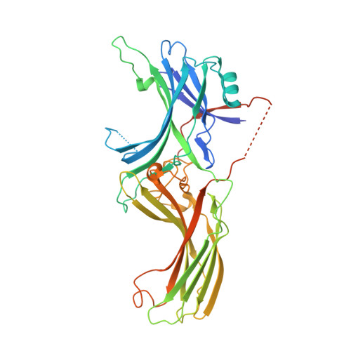
Structural evidence for visual arrestin priming via complexation of phosphoinositols.

Sander, C.L.Luu, J.Kim, K.Furkert, D.Jang, K.Reichenwallner, J.Kang, M.Lee, H.J.Eger, B.T.Choe, H.W.Fiedler, D.Ernst, O.P.Kim, Y.J.Palczewski, K.Kiser, P.D.

(2022) Structure 30: 263-277.e5

  • DOI: https://doi.org/10.1016/j.str.2021.10.002
  • Primary Citation of Related Structures:  
    7F1W, 7F1X, 7JSM, 7JTB, 7JXA, 7MOR, 7MP0, 7MP1, 7MP2

  • PubMed Abstract: 

    Visual arrestin (Arr1) terminates rhodopsin signaling by blocking its interaction with transducin. To do this, Arr1 translocates from the inner to the outer segment of photoreceptors upon light stimulation. Mounting evidence indicates that inositol phosphates (InsPs) affect Arr1 activity, but the Arr1-InsP molecular interaction remains poorly defined. We report the structure of bovine Arr1 in a ligand-free state featuring a near-complete model of the previously unresolved C-tail, which plays a crucial role in regulating Arr1 activity. InsPs bind to the N-domain basic patch thus displacing the C-tail, suggesting that they prime Arr1 for interaction with rhodopsin and help direct Arr1 translocation. These structures exhibit intact polar cores, suggesting that C-tail removal by InsP binding is insufficient to activate Arr1. These results show how Arr1 activity can be controlled by endogenous InsPs in molecular detail.


  • Organizational Affiliation

    Department of Pharmacology, Case Western Reserve University, Cleveland, OH 44106, USA; Department of Ophthalmology and the Gavin Herbert Eye Institute, University of California, Irvine, CA 92697, USA.


Macromolecules
Find similar proteins by:  (by identity cutoff)  |  3D Structure
Entity ID: 1
MoleculeChains Sequence LengthOrganismDetailsImage
S-arrestin
A, B, C, D
404Bos taurusMutation(s): 0 
UniProt
Find proteins for P08168 (Bos taurus)
Explore P08168 
Go to UniProtKB:  P08168
Entity Groups  
Sequence Clusters30% Identity50% Identity70% Identity90% Identity95% Identity100% Identity
UniProt GroupP08168
Sequence Annotations
Expand
  • Reference Sequence
Small Molecules
Ligands 3 Unique
IDChains Name / Formula / InChI Key2D Diagram3D Interactions
I3P (Subject of Investigation/LOI)
Query on I3P

Download Ideal Coordinates CCD File 
E [auth A],
G [auth B],
I [auth C],
N [auth D]
D-MYO-INOSITOL-1,4,5-TRIPHOSPHATE
C6 H15 O15 P3
MMWCIQZXVOZEGG-XJTPDSDZSA-N
ETX
Query on ETX

Download Ideal Coordinates CCD File 
J [auth C],
K [auth C],
L [auth C]
2-ETHOXYETHANOL
C4 H10 O2
ZNQVEEAIQZEUHB-UHFFFAOYSA-N
NA
Query on NA

Download Ideal Coordinates CCD File 
F [auth A],
H [auth B],
M [auth C]
SODIUM ION
Na
FKNQFGJONOIPTF-UHFFFAOYSA-N
Experimental Data & Validation

Experimental Data

  • Method: X-RAY DIFFRACTION
  • Resolution: 2.40 Å
  • R-Value Free: 0.249 
  • R-Value Work: 0.210 
  • R-Value Observed: 0.212 
  • Space Group: P 21 21 2
Unit Cell:
Length ( Å )Angle ( ˚ )
a = 167.782α = 90
b = 187.278β = 90
c = 90.54γ = 90
Software Package:
Software NamePurpose
REFMACrefinement
PDB_EXTRACTdata extraction
XDSdata reduction
XDSdata scaling
REFMACphasing

Structure Validation

View Full Validation Report


Ligand Structure Quality Assessment 


Entry History & Funding Information

Deposition Data


Funding OrganizationLocationGrant Number
National Institutes of Health/National Eye Institute (NIH/NEI)United StatesEY009339

Revision History  (Full details and data files)

  • Version 1.0: 2021-10-13
    Type: Initial release
  • Version 1.1: 2021-11-03
    Changes: Database references
  • Version 1.2: 2022-02-16
    Changes: Database references