7JWX

Crystal Structure of Trypsin Bound O-methyl Benzamidine


Experimental Data Snapshot

  • Method: X-RAY DIFFRACTION
  • Resolution: 2.38 Å
  • R-Value Free: 0.255 
  • R-Value Work: 0.212 
  • R-Value Observed: 0.214 

wwPDB Validation   3D Report Full Report


Ligand Structure Quality Assessment 


This is version 1.4 of the entry. See complete history


Literature

Small molecule peptidomimetic trypsin inhibitors: validation of an EKO binding mode, but with a twist.

Lyu, R.L.Joy, S.Packianathan, C.Laganowsky, A.Burgess, K.

(2022) Org Biomol Chem 20: 2075-2080

  • DOI: https://doi.org/10.1039/d1ob02127c
  • Primary Citation of Related Structures:  
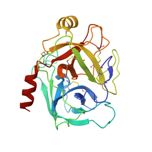
    7JWX

  • PubMed Abstract: 

    Examination of a series of naturally-occurring trypsin inhibitor proteins, led to identification of a set of three residues (which we call the "interface triplet") to be determinant of trypsin binding affinity, hence excellent templates for small molecule mimicry. Consequently, we attempted to use the Exploring Key Orientation (EKO) strategy developed in our lab to evaluate small molecules that mimic the interface triplet regions of natural trypsin inhibitors, and hence potentially might bind and inhibit the catalytic activity of trypsin. A bis-triazole scaffold ("TT-mer") was the most promising of the molecules evaluated in silico . Twelve such compounds were synthesized and assayed against trypsin, among which the best showed a K d of 2.1 μM. X-ray crystallography revealed a high degree of matching between an illustrative TT-mer's actual binding mode and that of the mimics that overlaid the interface triplet in the crystal structure. Deviation of the third side chain from the PPI structure seems to be due to alleviation of an unfavorable dipole-dipole interaction in the small molecule's actual bound conformation.


  • Organizational Affiliation

    Department of Chemistry, Texas A & M University, Box 30012, College Station, TX 77842-3012, USA. burgess@tamu.edu.


Macromolecules
Find similar proteins by:  (by identity cutoff)  |  3D Structure
Entity ID: 1
MoleculeChains Sequence LengthOrganismDetailsImage
Cationic trypsin
A, B, C, D
223Bos taurusMutation(s): 0 
EC: 3.4.21.4
UniProt
Find proteins for P00760 (Bos taurus)
Explore P00760 
Go to UniProtKB:  P00760
Entity Groups  
Sequence Clusters30% Identity50% Identity70% Identity90% Identity95% Identity100% Identity
UniProt GroupP00760
Sequence Annotations
Expand
  • Reference Sequence
Small Molecules
Ligands 5 Unique
IDChains Name / Formula / InChI Key2D Diagram3D Interactions
VN1
Query on VN1

Download Ideal Coordinates CCD File 
E [auth A],
H [auth B],
K [auth C],
O [auth D]
4-[(1-{(1S,2S)-1-[1-(4-aminobutyl)-1H-1,2,3-triazol-4-yl]-2-methylbutyl}-1H-1,2,3-triazol-4-yl)methoxy]-3-methoxybenzene-1-carboximidamide
C22 H33 N9 O2
DNIWQYWTKVVYDZ-BTYIYWSLSA-N
SO4
Query on SO4

Download Ideal Coordinates CCD File 
G [auth A],
M [auth C],
N [auth C],
R [auth D]
SULFATE ION
O4 S
QAOWNCQODCNURD-UHFFFAOYSA-L
GOL
Query on GOL

Download Ideal Coordinates CCD File 
P [auth D]GLYCEROL
C3 H8 O3
PEDCQBHIVMGVHV-UHFFFAOYSA-N
CA
Query on CA

Download Ideal Coordinates CCD File 
F [auth A],
I [auth B],
L [auth C],
Q [auth D]
CALCIUM ION
Ca
BHPQYMZQTOCNFJ-UHFFFAOYSA-N
CL
Query on CL

Download Ideal Coordinates CCD File 
J [auth B],
S [auth D]
CHLORIDE ION
Cl
VEXZGXHMUGYJMC-UHFFFAOYSA-M
Experimental Data & Validation

Experimental Data

  • Method: X-RAY DIFFRACTION
  • Resolution: 2.38 Å
  • R-Value Free: 0.255 
  • R-Value Work: 0.212 
  • R-Value Observed: 0.214 
  • Space Group: C 1 2 1
Unit Cell:
Length ( Å )Angle ( ˚ )
a = 189.27α = 90
b = 70.32β = 133
c = 137.74γ = 90
Software Package:
Software NamePurpose
PHENIXrefinement
PDB_EXTRACTdata extraction
XDSdata reduction
XSCALEdata scaling
PHASERphasing

Structure Validation

View Full Validation Report


Ligand Structure Quality Assessment 


Entry History 

Deposition Data

Revision History  (Full details and data files)

  • Version 1.0: 2021-08-04
    Type: Initial release
  • Version 1.1: 2022-02-23
    Changes: Database references
  • Version 1.2: 2022-03-16
    Changes: Database references
  • Version 1.3: 2022-03-23
    Changes: Database references
  • Version 1.4: 2023-10-18
    Changes: Data collection, Refinement description