7JWR

Cellular retinol-binding protein 2 (CRBP2) in complex with 2-oleoylglycerol


Experimental Data Snapshot

  • Method: X-RAY DIFFRACTION
  • Resolution: 1.30 Å
  • R-Value Free: 0.174 
  • R-Value Work: 0.134 
  • R-Value Observed: 0.136 

wwPDB Validation   3D Report Full Report


Ligand Structure Quality Assessment 


This is version 1.2 of the entry. See complete history


Literature

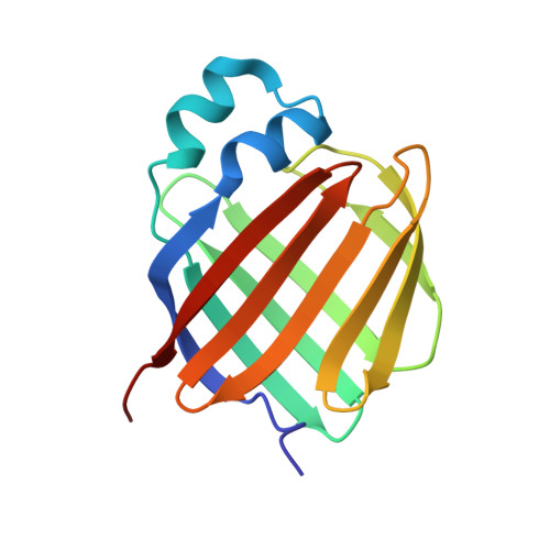
Molecular basis for the interaction of cellular retinol binding protein 2 (CRBP2) with nonretinoid ligands.

Silvaroli, J.A.Plau, J.Adams, C.H.Banerjee, S.Widjaja-Adhi, M.A.K.Blaner, W.S.Golczak, M.

(2021) J Lipid Res 62: 100054-100054

  • DOI: https://doi.org/10.1016/j.jlr.2021.100054
  • Primary Citation of Related Structures:  
    7JVG, 7JVY, 7JWD, 7JWR, 7JX2, 7JZ5, 7K3I

  • PubMed Abstract: 

    Present in the small intestine, cellular retinol binding protein 2 (CRBP2) plays an important role in the uptake, transport, and metabolism of dietary retinoids. However, the recent discovery of the interactions of CRBP2 with 2-arachidonoylglycerol and other monoacylglycerols (MAGs) suggests the broader involvement of this protein in lipid metabolism and signaling. To better understand the physiological role of CRBP2, we determined its protein-lipid interactome using a fluorescence-based retinol replacement assay adapted for a high-throughput screening format. By examining chemical libraries of bioactive lipids, we provided evidence for the selective interaction of CRBP2 with a subset of nonretinoid ligands with the highest affinity for sn-1 and sn-2 MAGs that contain polyunsaturated C18-C20 acyl chains. We also elucidated the structure-affinity relationship for nonretinoid ligands of this protein. We further dissect the molecular basis for this ligand's specificity by analyzing high-resolution crystal structures of CRBP2 in complex with selected derivatives of MAGs. Finally, we identify T51 and V62 as key amino acids that enable the broadening of ligand selectivity to MAGs in CRBP2 as compared with retinoid-specific CRBP1. Thus, our study provides the molecular framework for understanding the lipid selectivity and diverse functions of CRBPs in controlling lipid homeostasis.


  • Organizational Affiliation

    Department of Pharmacology, School of Medicine, Case Western Reserve University, Cleveland, OH, USA.


Macromolecules
Find similar proteins by:  (by identity cutoff)  |  3D Structure
Entity ID: 1
MoleculeChains Sequence LengthOrganismDetailsImage
Retinol-binding protein 2
A, B
138Homo sapiensMutation(s): 0 
Gene Names: RBP2CRBP2
UniProt & NIH Common Fund Data Resources
Find proteins for P50120 (Homo sapiens)
Explore P50120 
Go to UniProtKB:  P50120
PHAROS:  P50120
GTEx:  ENSG00000114113 
Entity Groups  
Sequence Clusters30% Identity50% Identity70% Identity90% Identity95% Identity100% Identity
UniProt GroupP50120
Sequence Annotations
Expand
  • Reference Sequence
Small Molecules
Experimental Data & Validation

Experimental Data

  • Method: X-RAY DIFFRACTION
  • Resolution: 1.30 Å
  • R-Value Free: 0.174 
  • R-Value Work: 0.134 
  • R-Value Observed: 0.136 
  • Space Group: P 21 21 21
Unit Cell:
Length ( Å )Angle ( ˚ )
a = 58.099α = 90
b = 67.38β = 90
c = 87.999γ = 90
Software Package:
Software NamePurpose
PHENIXrefinement
iMOSFLMdata reduction
Aimlessdata scaling
PHASERphasing

Structure Validation

View Full Validation Report


Ligand Structure Quality Assessment 


Entry History & Funding Information

Deposition Data


Funding OrganizationLocationGrant Number
National Institutes of Health/National Institute of Diabetes and Digestive and Kidney Disease (NIH/NIDDK)United StatesDK122071
National Institutes of Health/National Eye Institute (NIH/NEI)United StatesEY023948

Revision History  (Full details and data files)

  • Version 1.0: 2021-03-10
    Type: Initial release
  • Version 1.1: 2021-04-07
    Changes: Database references
  • Version 1.2: 2023-10-18
    Changes: Data collection, Database references, Refinement description