7JWI

Crystal structure of B17.R2 TCR in complex with H2D-b-NP366


Experimental Data Snapshot

  • Method: X-RAY DIFFRACTION
  • Resolution: 3.02 Å
  • R-Value Free: 0.281 
  • R-Value Work: 0.239 

wwPDB Validation   3D Report Full Report


This is version 1.1 of the entry. See complete history


Literature

Canonical T cell receptor docking on peptide-MHC is essential for T cell signaling.

Zareie, P.Szeto, C.Farenc, C.Gunasinghe, S.D.Kolawole, E.M.Nguyen, A.Blyth, C.Sng, X.Y.X.Li, J.Jones, C.M.Fulcher, A.J.Jacobs, J.R.Wei, Q.Wojciech, L.Petersen, J.Gascoigne, N.R.J.Evavold, B.D.Gaus, K.Gras, S.Rossjohn, J.La Gruta, N.L.

(2021) Science 372

  • DOI: https://doi.org/10.1126/science.abe9124
  • Primary Citation of Related Structures:  
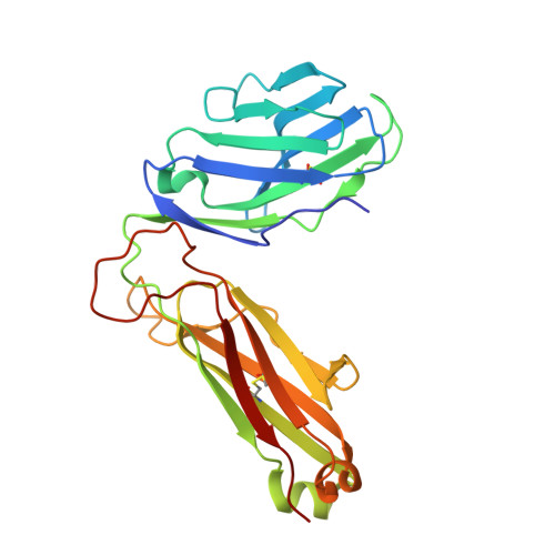
    7JWI, 7JWJ

  • PubMed Abstract: 

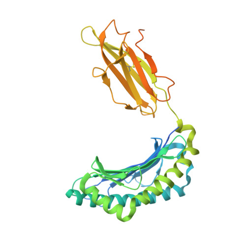
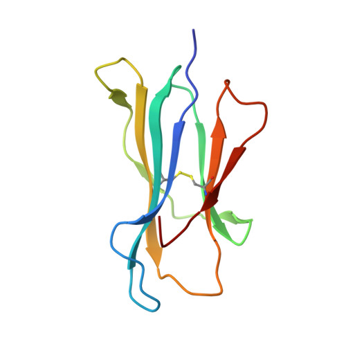
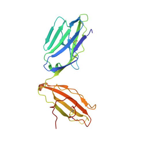
    T cell receptor (TCR) recognition of peptide-major histocompatibility complexes (pMHCs) is characterized by a highly conserved docking polarity. Whether this polarity is driven by recognition or signaling constraints remains unclear. Using "reversed-docking" TCRβ-variable (TRBV) 17 + TCRs from the naïve mouse CD8 + T cell repertoire that recognizes the H-2D b -NP 366 epitope, we demonstrate that their inability to support T cell activation and in vivo recruitment is a direct consequence of reversed docking polarity and not TCR-pMHCI binding or clustering characteristics. Canonical TCR-pMHCI docking optimally localizes CD8/Lck to the CD3 complex, which is prevented by reversed TCR-pMHCI polarity. The requirement for canonical docking was circumvented by dissociating Lck from CD8. Thus, the consensus TCR-pMHC docking topology is mandated by T cell signaling constraints.


  • Organizational Affiliation

    Infection and Immunity Program and Department of Biochemistry and Molecular Biology, Biomedicine Discovery Institute, Monash University, Clayton, Victoria, Australia.


Macromolecules
Find similar proteins by:  (by identity cutoff)  |  3D Structure
Entity ID: 1
MoleculeChains Sequence LengthOrganismDetailsImage
H-2 class I histocompatibility antigen, D-B alpha chain362Mus musculusMutation(s): 0 
Gene Names: H2-D1
UniProt
Find proteins for P01899 (Mus musculus)
Explore P01899 
Go to UniProtKB:  P01899
Entity Groups  
Sequence Clusters30% Identity50% Identity70% Identity90% Identity95% Identity100% Identity
UniProt GroupP01899
Sequence Annotations
Expand
  • Reference Sequence
Find similar proteins by:  (by identity cutoff)  |  3D Structure
Entity ID: 2
MoleculeChains Sequence LengthOrganismDetailsImage
Beta-2-microglobulin100Homo sapiensMutation(s): 0 
Gene Names: B2MCDABP0092HDCMA22P
UniProt & NIH Common Fund Data Resources
Find proteins for P61769 (Homo sapiens)
Explore P61769 
Go to UniProtKB:  P61769
PHAROS:  P61769
GTEx:  ENSG00000166710 
Entity Groups  
Sequence Clusters30% Identity50% Identity70% Identity90% Identity95% Identity100% Identity
UniProt GroupP61769
Sequence Annotations
Expand
  • Reference Sequence

Find similar proteins by:  Sequence   |   3D Structure  

Entity ID: 3
MoleculeChains Sequence LengthOrganismDetailsImage
Nucleoprotein9Influenza A virusMutation(s): 0 
UniProt
Find proteins for Q9Q0U8 (Influenza A virus (strain A/Goose/Guangdong/1/1996 H5N1 genotype Gs/Gd))
Explore Q9Q0U8 
Go to UniProtKB:  Q9Q0U8
Entity Groups  
UniProt GroupQ9Q0U8
Sequence Annotations
Expand
  • Reference Sequence
Find similar proteins by:  (by identity cutoff)  |  3D Structure
Entity ID: 4
MoleculeChains Sequence LengthOrganismDetailsImage
B17.R2 TCR alpha chain207Mus musculusMutation(s): 0 
Entity Groups  
Sequence Clusters30% Identity50% Identity70% Identity90% Identity95% Identity100% Identity
Sequence Annotations
Expand
  • Reference Sequence
Find similar proteins by:  (by identity cutoff)  |  3D Structure
Entity ID: 5
MoleculeChains Sequence LengthOrganismDetailsImage
B17.R2 TCR beta chain243Mus musculusMutation(s): 0 
Entity Groups  
Sequence Clusters30% Identity50% Identity70% Identity90% Identity95% Identity100% Identity
Sequence Annotations
Expand
  • Reference Sequence
Small Molecules
Ligands 1 Unique
IDChains Name / Formula / InChI Key2D Diagram3D Interactions
EDO
Query on EDO

Download Ideal Coordinates CCD File 
F [auth E]1,2-ETHANEDIOL
C2 H6 O2
LYCAIKOWRPUZTN-UHFFFAOYSA-N
Experimental Data & Validation

Experimental Data

  • Method: X-RAY DIFFRACTION
  • Resolution: 3.02 Å
  • R-Value Free: 0.281 
  • R-Value Work: 0.239 
  • Space Group: I 2 2 2
Unit Cell:
Length ( Å )Angle ( ˚ )
a = 91.668α = 90
b = 140.997β = 90
c = 217.649γ = 90
Software Package:
Software NamePurpose
BUSTERrefinement
PDB_EXTRACTdata extraction
XFITdata reduction
Aimlessdata scaling
PHASERphasing

Structure Validation

View Full Validation Report


Entry History 

Deposition Data

Revision History  (Full details and data files)

  • Version 1.0: 2021-07-07
    Type: Initial release
  • Version 1.1: 2023-10-18
    Changes: Data collection, Database references, Refinement description