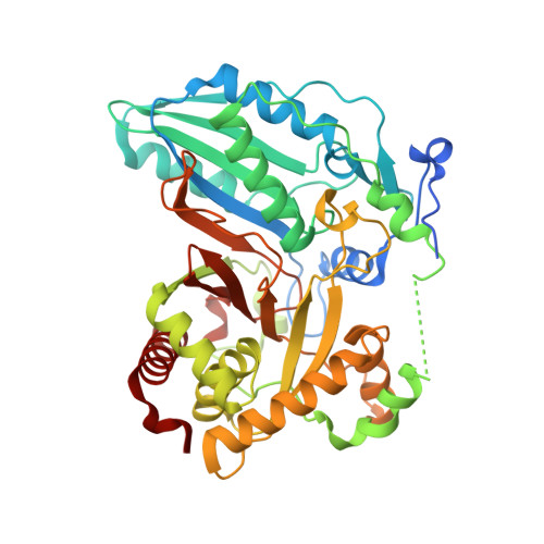
High-resolution structures of a siderophore-producing cyclization domain from Yersinia pestis offer a refined proposal of substrate binding.
Gnann, A.D., Xia, Y., Soule, J., Barthelemy, C., Mawani, J.S., Musoke, S.N., Castellano, B.M., Brignole, E.J., Frueh, D.P., Dowling, D.P.(2022) J Biol Chem 298: 102454-102454
- PubMed: 36063993
- DOI: https://doi.org/10.1016/j.jbc.2022.102454
- Primary Citation of Related Structures:
7JTJ, 7JUA - PubMed Abstract:
Nonribosomal peptide synthetase heterocyclization (Cy) domains generate biologically important oxazoline/thiazoline groups found in natural products, including pharmaceuticals and virulence factors such as some siderophores. Cy domains catalyze consecutive condensation and cyclodehydration reactions, although the mechanism is unknown. To better understand Cy domain catalysis, here we report the crystal structure of the second Cy domain (Cy2) of yersiniabactin synthetase from the causative agent of the plague, Yersinia pestis. Our high-resolution structure of Cy2 adopts a conformation that enables exploration of interactions with the extended thiazoline-containing cyclodehydration intermediate and the acceptor carrier protein (CP) to which it is tethered. We also report complementary electrostatic interfaces between Cy2 and its donor CP that mediate donor binding. Finally, we explored domain flexibility through normal mode analysis and identified small-molecule fragment-binding sites that may inform future antibiotic design targeting Cy function. Our results suggest how CP binding may influence global Cy conformations, with consequences for active-site remodeling to facilitate the separate condensation and cyclodehydration steps as well as potential inhibitor development.
Organizational Affiliation:
Department of Chemistry, University of Massachusetts Boston, Boston, Massachusetts, USA.