7JTI

Interphotoreceptor retinoid-binding protein (IRBP) in complex with a monoclonal antibody (F3F5 mAb5)


Experimental Data Snapshot

  • Method: ELECTRON MICROSCOPY
  • Resolution: 7.40 Å
  • Aggregation State: PARTICLE 
  • Reconstruction Method: SINGLE PARTICLE 

wwPDB Validation   3D Report Full Report


This is version 1.1 of the entry. See complete history


Literature

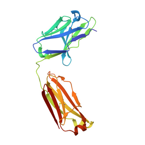
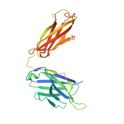
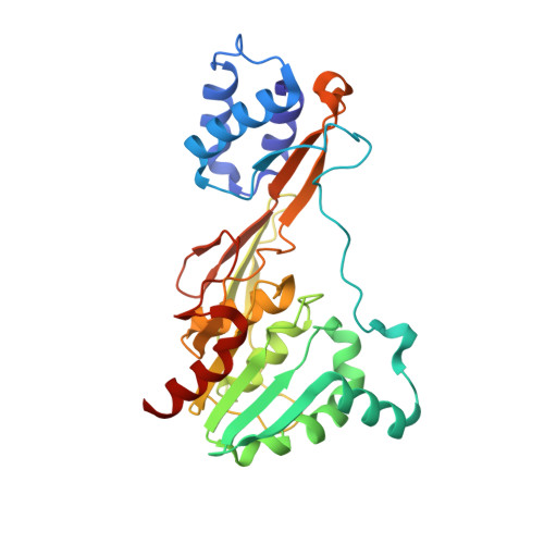
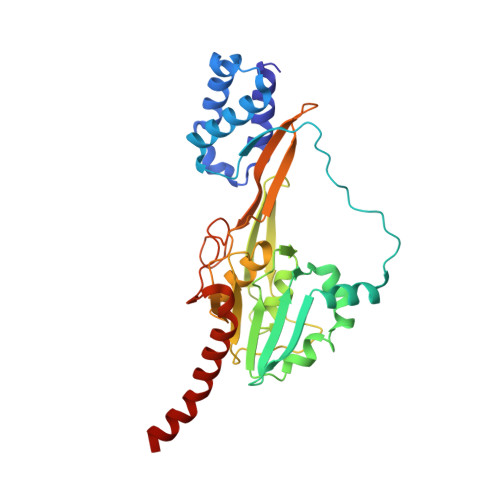
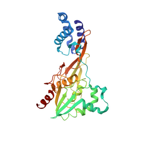
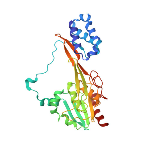
Single particle cryo-EM of the complex between interphotoreceptor retinoid-binding protein and a monoclonal antibody.

Sears, A.E.Albiez, S.Gulati, S.Wang, B.Kiser, P.Kovacik, L.Engel, A.Stahlberg, H.Palczewski, K.

(2020) FASEB J 34: 13918-13934

  • DOI: https://doi.org/10.1096/fj.202000796RR
  • Primary Citation of Related Structures:  
    7JTI

  • PubMed Abstract: 

    Interphotoreceptor retinoid-binding protein (IRBP) is a highly expressed protein secreted by rod and cone photoreceptors that has major roles in photoreceptor homeostasis as well as retinoid and polyunsaturated fatty acid transport between the neural retina and retinal pigment epithelium. Despite two crystal structures reported on fragments of IRBP and decades of research, the overall structure of IRBP and function within the visual cycle remain unsolved. Here, we studied the structure of native bovine IRBP in complex with a monoclonal antibody (mAb5) by cryo-electron microscopy, revealing the tertiary and quaternary structure at sufficient resolution to clearly identify the complex components. Complementary mass spectrometry experiments revealed the structure and locations of N-linked carbohydrate post-translational modifications. This work provides insight into the structure of IRBP, displaying an elongated, flexible three-dimensional architecture not seen among other retinoid-binding proteins. This work is the first step in elucidation of the function of this enigmatic protein.


  • Organizational Affiliation

    Department of Pharmacology, School of Medicine, Case Western Reserve University, Cleveland, OH, USA.


Macromolecules
Find similar proteins by:  (by identity cutoff)  |  3D Structure
Entity ID: 1
MoleculeChains Sequence LengthOrganismDetailsImage
mAb5 Fab light chainA [auth L]210Mus musculusMutation(s): 0 
Entity Groups  
Sequence Clusters30% Identity50% Identity70% Identity90% Identity95% Identity100% Identity
Sequence Annotations
Expand
  • Reference Sequence
Find similar proteins by:  (by identity cutoff)  |  3D Structure
Entity ID: 2
MoleculeChains Sequence LengthOrganismDetailsImage
mAb5 Fab heavy chainB [auth H]221Mus musculusMutation(s): 0 
Entity Groups  
Sequence Clusters30% Identity50% Identity70% Identity90% Identity95% Identity100% Identity
Sequence Annotations
Expand
  • Reference Sequence
Find similar proteins by:  (by identity cutoff)  |  3D Structure
Entity ID: 3
MoleculeChains Sequence LengthOrganismDetailsImage
Retinol-binding protein 3C [auth A]303Bos taurusMutation(s): 0 
UniProt
Find proteins for P12661 (Bos taurus)
Explore P12661 
Go to UniProtKB:  P12661
Entity Groups  
Sequence Clusters30% Identity50% Identity70% Identity90% Identity95% Identity100% Identity
UniProt GroupP12661
Sequence Annotations
Expand
  • Reference Sequence
Find similar proteins by:  (by identity cutoff)  |  3D Structure
Entity ID: 4
MoleculeChains Sequence LengthOrganismDetailsImage
Retinol-binding protein 3D [auth B]309Bos taurusMutation(s): 0 
UniProt
Find proteins for F1Q9N9 (Danio rerio)
Explore F1Q9N9 
Go to UniProtKB:  F1Q9N9
Entity Groups  
Sequence Clusters30% Identity50% Identity70% Identity90% Identity95% Identity100% Identity
UniProt GroupF1Q9N9
Sequence Annotations
Expand
  • Reference Sequence
Find similar proteins by:  (by identity cutoff)  |  3D Structure
Entity ID: 5
MoleculeChains Sequence LengthOrganismDetailsImage
Retinol-binding protein 3E [auth C]300Bos taurusMutation(s): 0 
UniProt
Find proteins for P12661 (Bos taurus)
Explore P12661 
Go to UniProtKB:  P12661
Entity Groups  
Sequence Clusters30% Identity50% Identity70% Identity90% Identity95% Identity100% Identity
UniProt GroupP12661
Sequence Annotations
Expand
  • Reference Sequence
Find similar proteins by:  (by identity cutoff)  |  3D Structure
Entity ID: 6
MoleculeChains Sequence LengthOrganismDetailsImage
Retinol-binding protein 3F [auth D]296Bos taurusMutation(s): 0 
UniProt
Find proteins for P12661 (Bos taurus)
Explore P12661 
Go to UniProtKB:  P12661
Entity Groups  
Sequence Clusters30% Identity50% Identity70% Identity90% Identity95% Identity100% Identity
UniProt GroupP12661
Sequence Annotations
Expand
  • Reference Sequence
Experimental Data & Validation

Experimental Data

  • Method: ELECTRON MICROSCOPY
  • Resolution: 7.40 Å
  • Aggregation State: PARTICLE 
  • Reconstruction Method: SINGLE PARTICLE 
EM Software:
TaskSoftware PackageVersion
RECONSTRUCTIONPHENIX
RECONSTRUCTIONUCSF Chimera
MODEL REFINEMENTUCSF Chimera

Structure Validation

View Full Validation Report


Entry History & Funding Information

Deposition Data


Funding OrganizationLocationGrant Number
National Institutes of Health/National Eye Institute (NIH/NEI)United StatesR24EY024864
National Institutes of Health/National Eye Institute (NIH/NEI)United StatesR24EY027283
National Institutes of Health/National Eye Institute (NIH/NEI)United StatesT32 GM002750
Swiss National Science FoundationSwitzerlandNCCR TransCure

Revision History  (Full details and data files)

  • Version 1.0: 2020-10-07
    Type: Initial release
  • Version 1.1: 2020-12-16
    Changes: Database references