Capsid Structure of Anabaena Cyanophage A-1(L).
Cui, N., Yang, F., Zhang, J.T., Sun, H., Chen, Y., Yu, R.C., Chen, Z.P., Jiang, Y.L., Han, S.J., Xu, X., Li, Q., Zhou, C.Z.(2021) J Virol 95: e0135621-e0135621
- PubMed: 34549983
- DOI: https://doi.org/10.1128/JVI.01356-21
- Primary Citation of Related Structures:
7F38 - PubMed Abstract:
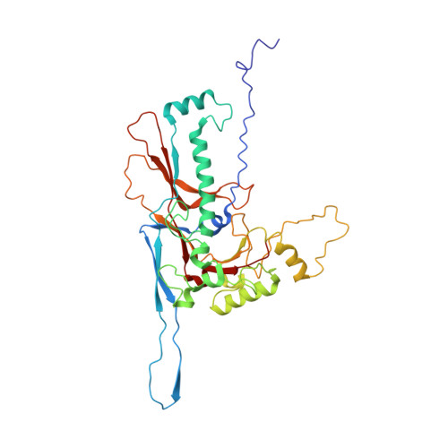
A-1(L) is a freshwater cyanophage with a contractile tail that specifically infects Anabaena sp. PCC 7120, one of the model strains for molecular studies of cyanobacteria. Although isolated for half a century, its structure remains unknown, which limits our understanding on the interplay between A-1(L) and its host. Here we report the 3.35 Å cryo-EM structure of A-1(L) capsid, representing the first near-atomic resolution structure of a phage capsid with a T number of 9. The major capsid gp4 proteins assemble into 91 capsomers, including 80 hexons: 20 at the center of the facet and 60 at the facet edge, in addition to 11 identical pentons. These capsomers further assemble into the icosahedral capsid, via gradually increasing curvatures. Different from the previously reported capsids of known-structure, A-1(L) adopts a noncovalent chainmail structure of capsid stabilized by two kinds of mortise-and-tenon inter-capsomer interactions: a three-layered interface at the pseudo 3-fold axis combined with the complementarity in shape and electrostatic potential around the 2-fold axis. This unique capsomer construction enables A-1(L) to possess a rigid capsid, which is solely composed of the major capsid proteins with an HK97 fold. IMPORTANCE Cyanobacteria are the most abundant photosynthetic bacteria, contributing significantly to the biomass production, O 2 generation, and CO 2 consumption on our planet. Their community structure and homeostasis in natural aquatic ecosystems are largely regulated by the corresponding cyanophages. In this study, we solved the structure of cyanophage A-1(L) capsid at near-atomic resolution and revealed a unique capsid construction. This capsid structure provides the molecular details for better understanding the assembly of A-1(L), and a structural platform for future investigation and application of A-1(L) in combination with its host Anabaena sp. PCC 7120. As the first isolated freshwater cyanophage that infects the genetically tractable model cyanobacterium, A-1(L) should become an ideal template for the genetic engineering and synthetic biology studies.
Organizational Affiliation:
Hefei National Laboratory for Physical Sciences at the Microscale and School of Life Sciences, University of Science and Technology of Chinagrid.59053.3a, Hefei, Anhui, China.