7EXZ

DgpB-DgpC complex apo 2.5 angstrom


Experimental Data Snapshot

  • Method: X-RAY DIFFRACTION
  • Resolution: 2.50 Å
  • R-Value Free: 0.233 
  • R-Value Work: 0.182 
  • R-Value Observed: 0.183 

wwPDB Validation   3D Report Full Report


This is version 1.2 of the entry. See complete history


Literature

C-Glycoside metabolism in the gut and in nature: Identification, characterization, structural analyses and distribution of C-C bond-cleaving enzymes.

Mori, T.Kumano, T.He, H.Watanabe, S.Senda, M.Moriya, T.Adachi, N.Hori, S.Terashita, Y.Kawasaki, M.Hashimoto, Y.Awakawa, T.Senda, T.Abe, I.Kobayashi, M.

(2021) Nat Commun 12: 6294-6294

  • DOI: https://doi.org/10.1038/s41467-021-26585-1
  • Primary Citation of Related Structures:  
    7BVR, 7BVS, 7DRD, 7DRE, 7EXB, 7EXZ

  • PubMed Abstract: 

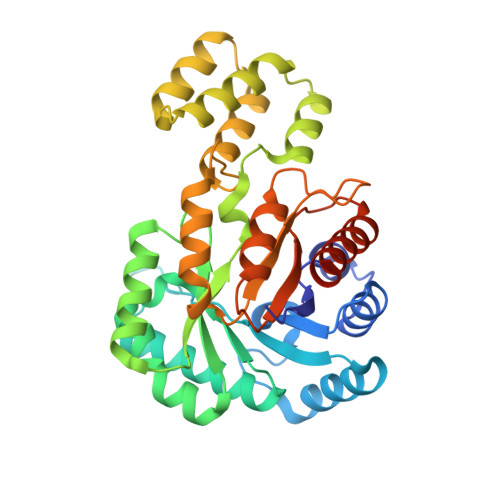
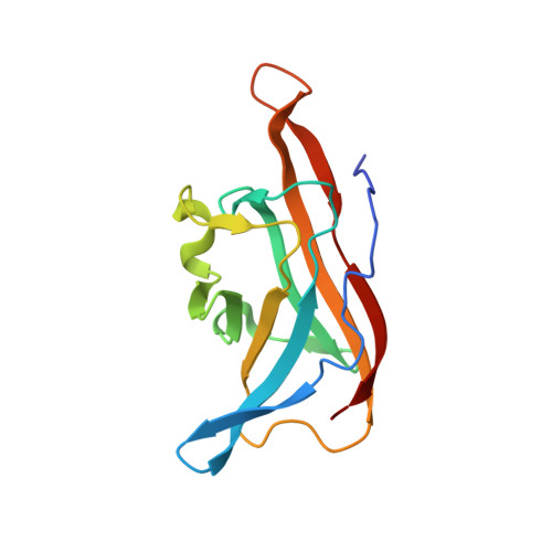
    C-Glycosides, in which a sugar moiety is linked via a carbon-carbon (C-C) bond to a non-sugar moiety (aglycone), are found in our food and medicine. The C-C bond is cleaved by intestinal microbes and the resulting aglycones exert various bioactivities. Although the enzymes responsible for the reactions have been identified, their catalytic mechanisms and the generality of the reactions in nature remain to be explored. Here, we present the identification and structural basis for the activation of xenobiotic C-glycosides by heterocomplex C-deglycosylation enzymes from intestinal and soil bacteria. They are found to be metal-dependent enzymes exhibiting broad substrate specificity toward C-glycosides. X-ray crystallographic and cryo-electron microscopic analyses, as well as structure-based mutagenesis, reveal the structural details of these enzymes and the detailed catalytic mechanisms of their remarkable C-C bond cleavage reactions. Furthermore, bioinformatic and biochemical analyses suggest that the C-deglycosylation enzymes are widely distributed in the gut, soil, and marine bacteria.


  • Organizational Affiliation

    Graduate School of Pharmaceutical Sciences, The University of Tokyo, 7-3-1 Hongo, Bunkyo-ku, Tokyo, 113-0033, Japan.


Macromolecules
Find similar proteins by:  (by identity cutoff)  |  3D Structure
Entity ID: 1
MoleculeChains Sequence LengthOrganismDetailsImage
AP_endonuc_2 domain-containing protein
A, C, E, G
337human intestinal bacterium PUEMutation(s): 0 
Gene Names: dgpC
UniProt
Find proteins for A0A3Q9WXL1 (human intestinal bacterium PUE)
Explore A0A3Q9WXL1 
Go to UniProtKB:  A0A3Q9WXL1
Entity Groups  
Sequence Clusters30% Identity50% Identity70% Identity90% Identity95% Identity100% Identity
UniProt GroupA0A3Q9WXL1
Sequence Annotations
Expand
  • Reference Sequence
Find similar proteins by:  (by identity cutoff)  |  3D Structure
Entity ID: 2
MoleculeChains Sequence LengthOrganismDetailsImage
DgpB
B, D, F, H
142human intestinal bacterium PUEMutation(s): 0 
Gene Names: dgpB
UniProt
Find proteins for A0A3Q9WUX0 (human intestinal bacterium PUE)
Explore A0A3Q9WUX0 
Go to UniProtKB:  A0A3Q9WUX0
Entity Groups  
Sequence Clusters30% Identity50% Identity70% Identity90% Identity95% Identity100% Identity
UniProt GroupA0A3Q9WUX0
Sequence Annotations
Expand
  • Reference Sequence
Small Molecules
Ligands 4 Unique
IDChains Name / Formula / InChI Key2D Diagram3D Interactions
EPE
Query on EPE

Download Ideal Coordinates CCD File 
IA [auth H],
L [auth B],
M [auth B],
T [auth D],
Z [auth F]
4-(2-HYDROXYETHYL)-1-PIPERAZINE ETHANESULFONIC ACID
C8 H18 N2 O4 S
JKMHFZQWWAIEOD-UHFFFAOYSA-N
SO4
Query on SO4

Download Ideal Coordinates CCD File 
AA [auth F]
BA [auth F]
EA [auth G]
FA [auth G]
GA [auth G]
AA [auth F],
BA [auth F],
EA [auth G],
FA [auth G],
GA [auth G],
HA [auth G],
JA [auth H],
K [auth A],
KA [auth H],
N [auth B],
O [auth B],
R [auth C],
S [auth C],
U [auth D],
V [auth D],
Y [auth E]
SULFATE ION
O4 S
QAOWNCQODCNURD-UHFFFAOYSA-L
GOL
Query on GOL

Download Ideal Coordinates CCD File 
DA [auth G],
J [auth A],
Q [auth C],
X [auth E]
GLYCEROL
C3 H8 O3
PEDCQBHIVMGVHV-UHFFFAOYSA-N
MN (Subject of Investigation/LOI)
Query on MN

Download Ideal Coordinates CCD File 
CA [auth G],
I [auth A],
P [auth C],
W [auth E]
MANGANESE (II) ION
Mn
WAEMQWOKJMHJLA-UHFFFAOYSA-N
Experimental Data & Validation

Experimental Data

  • Method: X-RAY DIFFRACTION
  • Resolution: 2.50 Å
  • R-Value Free: 0.233 
  • R-Value Work: 0.182 
  • R-Value Observed: 0.183 
  • Space Group: P 21 21 21
Unit Cell:
Length ( Å )Angle ( ˚ )
a = 86.516α = 90
b = 156.721β = 90
c = 158.931γ = 90
Software Package:
Software NamePurpose
PHENIXrefinement
XDSdata reduction
XSCALEdata scaling
PHASERphasing

Structure Validation

View Full Validation Report


Entry History 

Deposition Data

Revision History  (Full details and data files)

  • Version 1.0: 2021-11-03
    Type: Initial release
  • Version 1.1: 2021-11-17
    Changes: Database references
  • Version 1.2: 2023-11-29
    Changes: Data collection, Refinement description