7EVS

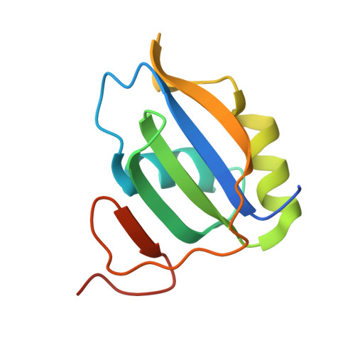
Crystal structure of hnRNP LL RRM2 in complex with SETD2


Experimental Data Snapshot

  • Method: X-RAY DIFFRACTION
  • Resolution: 1.60 Å
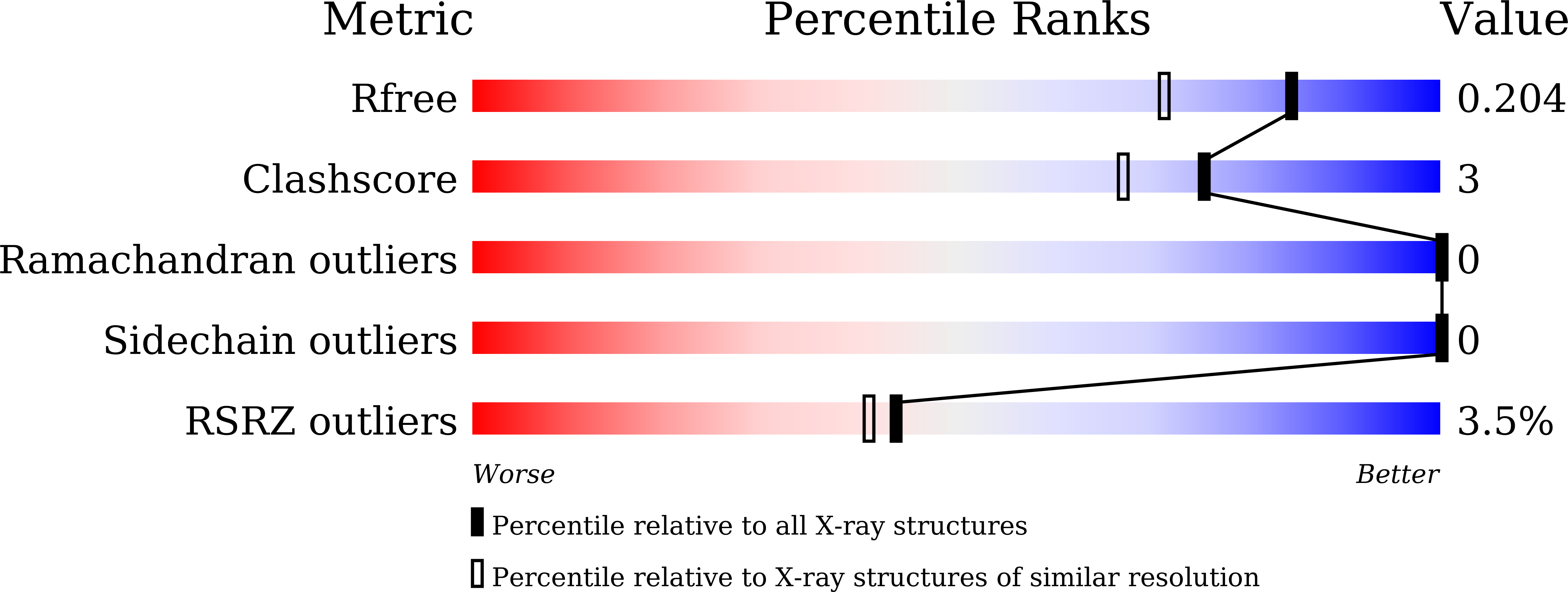
  • R-Value Free: 0.204 
  • R-Value Work: 0.178 
  • R-Value Observed: 0.179 

wwPDB Validation   3D Report Full Report


This is version 1.2 of the entry. See complete history


Literature

Structural basis of the interaction between SETD2 methyltransferase and hnRNP L paralogs for governing co-transcriptional splicing.

Bhattacharya, S.Wang, S.Reddy, D.Shen, S.Zhang, Y.Zhang, N.Li, H.Washburn, M.P.Florens, L.Shi, Y.Workman, J.L.Li, F.

(2021) Nat Commun 12: 6452-6452

  • DOI: https://doi.org/10.1038/s41467-021-26799-3
  • Primary Citation of Related Structures:  
    7EVR, 7EVS

  • PubMed Abstract: 

    The RNA recognition motif (RRM) binds to nucleic acids as well as proteins. More than one such domain is found in the pre-mRNA processing hnRNP proteins. While the mode of RNA recognition by RRMs is known, the molecular basis of their protein interaction remains obscure. Here we describe the mode of interaction between hnRNP L and LL with the methyltransferase SETD2. We demonstrate that for the interaction to occur, a leucine pair within a highly conserved stretch of SETD2 insert their side chains in hydrophobic pockets formed by hnRNP L RRM2. Notably, the structure also highlights that RRM2 can form a ternary complex with SETD2 and RNA. Remarkably, mutating the leucine pair in SETD2 also results in its reduced interaction with other hnRNPs. Importantly, the similarity that the mode of SETD2-hnRNP L interaction shares with other related protein-protein interactions reveals a conserved design by which splicing regulators interact with one another.


  • Organizational Affiliation

    Stowers Institute for Medical Research, Kansas City, MO, 64110, USA.


Macromolecules
Find similar proteins by:  (by identity cutoff)  |  3D Structure
Entity ID: 1
MoleculeChains Sequence LengthOrganismDetailsImage
Heterogeneous nuclear ribonucleoprotein L-like
A, B
110Homo sapiensMutation(s): 0 
Gene Names: HNRNPLLHNRPLLSRRFBLOCK24
UniProt & NIH Common Fund Data Resources
Find proteins for Q8WVV9 (Homo sapiens)
Explore Q8WVV9 
Go to UniProtKB:  Q8WVV9
PHAROS:  Q8WVV9
GTEx:  ENSG00000143889 
Entity Groups  
Sequence Clusters30% Identity50% Identity70% Identity90% Identity95% Identity100% Identity
UniProt GroupQ8WVV9
Sequence Annotations
Expand
  • Reference Sequence

Find similar proteins by:  Sequence   |   3D Structure  

Entity ID: 2
MoleculeChains Sequence LengthOrganismDetailsImage
SHI domain from Histone-lysine N-methyltransferase SETD2
C, D
13Homo sapiensMutation(s): 0 
UniProt & NIH Common Fund Data Resources
Find proteins for Q9BYW2 (Homo sapiens)
Explore Q9BYW2 
Go to UniProtKB:  Q9BYW2
PHAROS:  Q9BYW2
GTEx:  ENSG00000181555 
Entity Groups  
Sequence Clusters30% Identity50% Identity70% Identity90% Identity95% Identity100% Identity
UniProt GroupQ9BYW2
Sequence Annotations
Expand
  • Reference Sequence
Experimental Data & Validation

Experimental Data

  • Method: X-RAY DIFFRACTION
  • Resolution: 1.60 Å
  • R-Value Free: 0.204 
  • R-Value Work: 0.178 
  • R-Value Observed: 0.179 
  • Space Group: P 1
Unit Cell:
Length ( Å )Angle ( ˚ )
a = 41.676α = 85.02
b = 41.685β = 89.57
c = 42.636γ = 71.12
Software Package:
Software NamePurpose
HKL-2000data scaling
PHENIXrefinement
PDB_EXTRACTdata extraction
HKL-2000data reduction
MOLREPphasing

Structure Validation

View Full Validation Report


Entry History 

Deposition Data

Revision History  (Full details and data files)

  • Version 1.0: 2021-10-20
    Type: Initial release
  • Version 1.1: 2022-12-21
    Changes: Database references
  • Version 1.2: 2023-11-29
    Changes: Data collection, Refinement description