7EPU

Crystal structure of HsALC1


Experimental Data Snapshot

  • Method: X-RAY DIFFRACTION
  • Resolution: 3.50 Å
  • R-Value Free: 0.316 
  • R-Value Work: 0.268 
  • R-Value Observed: 0.272 

wwPDB Validation   3D Report Full Report


Ligand Structure Quality Assessment 


This is version 1.1 of the entry. See complete history


Literature

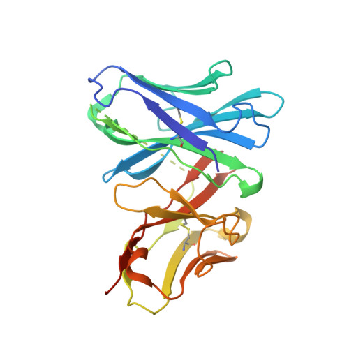
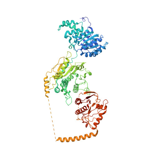
Structural basis of ALC1/CHD1L autoinhibition and the mechanism of activation by the nucleosome.

Wang, L.Chen, K.Chen, Z.

(2021) Nat Commun 12: 4057-4057

  • DOI: https://doi.org/10.1038/s41467-021-24320-4
  • Primary Citation of Related Structures:  
    7ENN, 7EPU

  • PubMed Abstract: 

    Chromatin remodeler ALC1 (amplification in liver cancer 1) is crucial for repairing damaged DNA. It is autoinhibited and activated by nucleosomal epitopes. However, the mechanisms by which ALC1 is regulated remain unclear. Here we report the crystal structure of human ALC1 and the cryoEM structure bound to the nucleosome. The structure shows the macro domain of ALC1 binds to lobe 2 of the ATPase motor, sequestering two elements for nucleosome recognition, explaining the autoinhibition mechanism of the enzyme. The H4 tail competes with the macro domain for lobe 2-binding, explaining the requirement for this nucleosomal epitope for ALC1 activation. A dual-arginine-anchor motif of ALC1 recognizes the acidic pocket of the nucleosome, which is critical for chromatin remodeling in vitro. Together, our findings illustrate the structures of ALC1 and shed light on its regulation mechanisms, paving the way for the discovery of drugs targeting ALC1 for the treatment of cancer.


  • Organizational Affiliation

    Key Laboratory for Protein Sciences of Ministry of Education, School of Life Science, Tsinghua University, 100084, Beijing, P.R. China.


Macromolecules
Find similar proteins by:  (by identity cutoff)  |  3D Structure
Entity ID: 1
MoleculeChains Sequence LengthOrganismDetailsImage
non-immunized human scFv263Homo sapiensMutation(s): 0 
Entity Groups  
Sequence Clusters30% Identity50% Identity70% Identity90% Identity95% Identity100% Identity
Sequence Annotations
Expand
  • Reference Sequence
Find similar proteins by:  (by identity cutoff)  |  3D Structure
Entity ID: 2
MoleculeChains Sequence LengthOrganismDetailsImage
Chromodomain-helicase-DNA-binding protein 1-like880Homo sapiensMutation(s): 0 
Gene Names: CHD1LALC1
EC: 3.6.4.12
UniProt & NIH Common Fund Data Resources
Find proteins for Q86WJ1 (Homo sapiens)
Explore Q86WJ1 
Go to UniProtKB:  Q86WJ1
PHAROS:  Q86WJ1
GTEx:  ENSG00000131778 
Entity Groups  
Sequence Clusters30% Identity50% Identity70% Identity90% Identity95% Identity100% Identity
UniProt GroupQ86WJ1
Sequence Annotations
Expand
  • Reference Sequence
Experimental Data & Validation

Experimental Data

  • Method: X-RAY DIFFRACTION
  • Resolution: 3.50 Å
  • R-Value Free: 0.316 
  • R-Value Work: 0.268 
  • R-Value Observed: 0.272 
  • Space Group: C 2 2 21
Unit Cell:
Length ( Å )Angle ( ˚ )
a = 153.48α = 90
b = 225.263β = 90
c = 106.533γ = 90
Software Package:
Software NamePurpose
PHENIXrefinement
HKL-2000data scaling
PDB_EXTRACTdata extraction
HKL-2000data reduction
PHASERphasing

Structure Validation

View Full Validation Report


Ligand Structure Quality Assessment 


Entry History 

Deposition Data

Revision History  (Full details and data files)

  • Version 1.0: 2021-07-14
    Type: Initial release
  • Version 1.1: 2023-11-29
    Changes: Data collection, Database references, Refinement description