7EPB

Cryo-EM structure of LY354740-bound mGlu2 homodimer


Experimental Data Snapshot

  • Method: ELECTRON MICROSCOPY
  • Resolution: 3.10 Å
  • Aggregation State: PARTICLE 
  • Reconstruction Method: SINGLE PARTICLE 

wwPDB Validation   3D Report Full Report


This is version 1.1 of the entry. See complete history


Literature

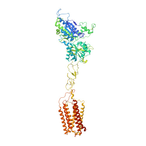
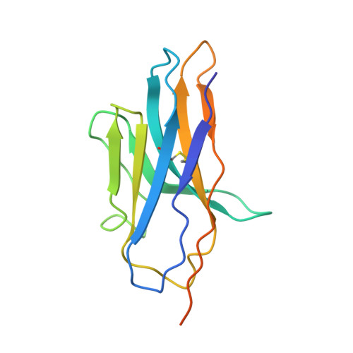
Structures of human mGlu2 and mGlu7 homo- and heterodimers.

Du, J.Wang, D.Fan, H.Xu, C.Tai, L.Lin, S.Han, S.Tan, Q.Wang, X.Xu, T.Zhang, H.Chu, X.Yi, C.Liu, P.Wang, X.Zhou, Y.Pin, J.P.Rondard, P.Liu, H.Liu, J.Sun, F.Wu, B.Zhao, Q.

(2021) Nature 594: 589-593

  • DOI: https://doi.org/10.1038/s41586-021-03641-w
  • Primary Citation of Related Structures:  
    7EPA, 7EPB, 7EPC, 7EPD, 7EPE, 7EPF

  • PubMed Abstract: 

    The metabotropic glutamate receptors (mGlus) are involved in the modulation of synaptic transmission and neuronal excitability in the central nervous system 1 . These receptors probably exist as both homo- and heterodimers that have unique pharmacological and functional properties 2-4 . Here we report four cryo-electron microscopy structures of the human mGlu subtypes mGlu2 and mGlu7, including inactive mGlu2 and mGlu7 homodimers; mGlu2 homodimer bound to an agonist and a positive allosteric modulator; and inactive mGlu2-mGlu7 heterodimer. We observed a subtype-dependent dimerization mode for these mGlus, as a unique dimer interface that is mediated by helix IV (and that is important for limiting receptor activity) exists only in the inactive mGlu2 structure. The structures provide molecular details of the inter- and intra-subunit conformational changes that are required for receptor activation, which distinguish class C G-protein-coupled receptors from those in classes A and B. Furthermore, our structure and functional studies of the mGlu2-mGlu7 heterodimer suggest that the mGlu7 subunit has a dominant role in controlling dimeric association and G-protein activation in the heterodimer. These insights into mGlu homo- and heterodimers highlight the complex landscape of mGlu dimerization and activation.


  • Organizational Affiliation

    State Key Laboratory of Drug Research, Shanghai Institute of Materia Medica, Chinese Academy of Sciences, Shanghai, China.


Macromolecules
Find similar proteins by:  (by identity cutoff)  |  3D Structure
Entity ID: 1
MoleculeChains Sequence LengthOrganismDetailsImage
Metabotropic glutamate receptor 2
A, B
851Homo sapiensMutation(s): 1 
Gene Names: GRM2GPRC1BMGLUR2
Membrane Entity: Yes 
UniProt & NIH Common Fund Data Resources
Find proteins for Q14416 (Homo sapiens)
Explore Q14416 
Go to UniProtKB:  Q14416
PHAROS:  Q14416
GTEx:  ENSG00000164082 
Entity Groups  
Sequence Clusters30% Identity50% Identity70% Identity90% Identity95% Identity100% Identity
UniProt GroupQ14416
Sequence Annotations
Expand
  • Reference Sequence
Find similar proteins by:  (by identity cutoff)  |  3D Structure
Entity ID: 2
MoleculeChains Sequence LengthOrganismDetailsImage
Anti-RON nanobody
C, D
137Lama glamaMutation(s): 0 
Entity Groups  
Sequence Clusters30% Identity50% Identity70% Identity90% Identity95% Identity100% Identity
Sequence Annotations
Expand
  • Reference Sequence
Small Molecules
Ligands 1 Unique
IDChains Name / Formula / InChI Key2D Diagram3D Interactions
40F (Subject of Investigation/LOI)
Query on 40F

Download Ideal Coordinates CCD File 
E [auth A],
F [auth B]
(1S,2S,5R,6S)-2-aminobicyclo[3.1.0]hexane-2,6-dicarboxylic acid
C8 H11 N O4
VTAARTQTOOYTES-RGDLXGNYSA-N
Experimental Data & Validation

Experimental Data

  • Method: ELECTRON MICROSCOPY
  • Resolution: 3.10 Å
  • Aggregation State: PARTICLE 
  • Reconstruction Method: SINGLE PARTICLE 

Structure Validation

View Full Validation Report


Entry History & Funding Information

Deposition Data


Funding OrganizationLocationGrant Number
National Science Foundation (NSF, China)China31825010
National Science Foundation (NSF, China)China81525024
National Science Foundation (NSF, China)China31830020
National Science Foundation (NSF, China)China81720108031

Revision History  (Full details and data files)

  • Version 1.0: 2021-06-23
    Type: Initial release
  • Version 1.1: 2021-07-07
    Changes: Database references